Biotensegrity-Informed Physical Exam
When he demonstrates the moment of impact, the diffusion of forces can be seen as his left bra- chium is driven into the ribs and the left scapula is driven medially wfith some glenoid depression and counter-clockwise rotation of the scapula. This leads to more detailed examination of the left flank, w'hich reveals taut bands and latent trigger points in the low'er latissimus dorsi (LD), low'er serratus anterior, and adjacent external oblique fibers at the low ribs. Focal pressure at the rib enthe- ses results in pain response as well. If his remote injury in this area did not fully repair, the connective tissue here would be lax. The taut muscle fibers in this region could be a response to connective tissue laxity. Could passive tension be returned to the system temporarily through more diffuse palpation? How would this change his examination? When support is applied to this region, with the intent of restoring some of the pre-stress lost in the accident, two remarkable changes occur:
- 1. The apprehension test now has full, painless PROM into abduction/external rotation (Figure 10.8).
- 2. Speed’s testing in supination and pronation now displays normal strength and stability (Figures 10.9 and 10.10).
Movement is not bending of hinges, but expansion, repositioning and contraction of tensegrities.
Levin and Martin20
With pre-stress added back into the system, the quality of active muscle contraction during Speed’s testing changes from an attempt at “pulling” the upper extremity into position to “an expansion” of the upper extremity and trunk into the applied downward force. The movement is now a “complex whole” rather than an uncoordinated contraction of parts (videos 7 and 8). To confirm these observations, the tests are repeated without the pre-stress applied to the left flank. They revert back to the uncoordinated contraction of parts and pain with limited PROM. Let’s explore these observations in the context of fascial anatomy and trigger point science.
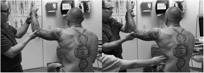
FIGURE 10.8 Biotensegrity-informed physical exam: left image same as Figure 10.3. Right image same test performed; however, prior to beginning the test, tension (pre-stress) is added via palpation pressure at an area of remote trauma. PROM normalizes, and he has no posterior pain (see video 6).
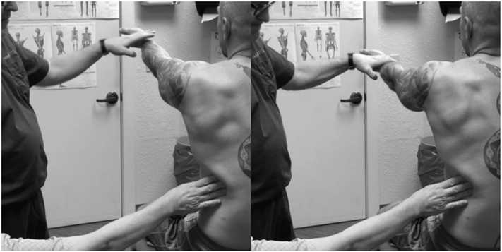
FIGURE 10.9 Biotensegrity-informed Speed’s test in supination: with the arm resting on his lap, pressure is applied at left low ribs/LD/SPI; then, test is repeated with full strength (see video 7).
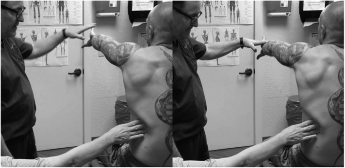
FIGURE 10.10 Biotensegrity-informed Speed’s test in pronation: with the arm resting on his lap, pressure is applied at left low ribs/LD/SPI; then, test is repeated with full strength (see video 8).

FIGURE 10.11 Ultrasound imaged uncompressed: probe is long axis on the ninth rib with three muscles visualized. Left image is unlabeled; right with muscles outlined LD in red, SA in blue, and EO in green.
The False Concept of Ligaments as the Only Controllers of PROM
In musculoskeletal anatomy, ligaments (including joint capsules) are conceived as the passive controllers of joint motion. Tests of ligamentous and capsular integrity focus on forces applied to a joint while the patient is relaxed, such as this apprehension test designed to evaluate joint stability. If there is posterior impingement with this test in an overhead athlete, the conceptual explanation then becomes laxity in the anterior capsule versus tightness in the posterior, inferior joint capsule which pushes the humeral head anterior, creating posterior impingement.21 Yet, in our patient, adding tension by applying pressure at the flank improves the passive dynamics at the shoulder joint.
Van Der Wal22 has argued that the radial collateral and annular ligaments of the lateral elbow do not exist as separate structures from the myofascial continuity of the extensor forearm mechanism. He argues that the appearance of a ligament structure in this location is an artifact of the dissection process. Ultimately, Van Der Wal argues that muscle tendon units are more commonly in series with ligamentous tissue rather than in parallel as most anatomic drawings illustrate. Numerous examples of muscle to tendon to joint capsule have been described in the knee,23 hip,24 and shoulder.25
The muscle-fascial continuity, then, is a pre-stressed controller of PROM, just as the ligaments and joint capsules have been described. In fact, Masi et al. have described the resting tone of muscles as a significant contributor to dynamic control.26 This broadly extended, impossibly complex continuity of tension has been described by Levin et al. as a multi-bar kinematic mechanism of biologic movement.27 So, what part of this mechanism was injured in our patient during the MVA? At the ninth rib, three muscles partially blend with each other: serratus anterior (SA), external oblique (EO), and latissimus dorsi (LD). Dynamic ultrasonography with compression at this location reveals the poor definition and instability of this blending (Figures 10.11 and 10.12). Serratus anterior translates toward the inferior angle of the scapula which reveals incompetence of the muscle as it blends with the periosteum of the rib and the adjoining external oblique. LD also compresses easily and

FIGURE 10.12 Ultrasound imaged with compression showing loss of tensional integrity (see video 9): probe is long axis on the ninth rib with three muscles visualized. Left image is unlabeled; right with muscles outlined LD in red, SA in blue, and EO in green.
translates toward the scapula (video 9). Similar weakness is seen at the serratus posterior inferior (SPI) and LD insertions at ribs 10 through 12.

