Al Types and Applications
Before we discuss the different types of Al and its applications, it is important to define what Al is? There are numerous definitions of Al in the literature, but this one derived from computer science describes Al as “the study of intelligent agents and systems, exhibiting the ability to accomplish complex goals” (AAIH, 2019)- However, this definition is oriented to an academic perspective. From an application and industry perspective, Al can be best described as “machines assuming intelligence”. Now that we have defined Al, it is pertinent to mention here two levels of AL General and Narrow Al. General Al, also referred to as Artificial General Intelligence, is when Al exhibits “a full range of cognitive abilities or general intelligence actions by an intelligent agent or system” (AAIH, 2019), while Narrow Al, also referred to as Weak Al, is where Al is specified to address a singular or limited task.

Figure 1.2 Al types, learning approaches and applications.
The predominant approach of Al, currently, is ML (Figure 1.2). This approach involves performing tasks without explicit instructions relying mainly on patterns and relationships in the training data and environment (AAIH, 2019). To develop ML models, you will need to define the necessary features, i.e. dependent and independent or input and target variables, and develop datasets including the features. Further to this, you split up the dataset into training and test datasets to allow for internal validation. Following this, the datasets are trained or tested with relevant ML algorithms. If the training dataset contains the input data and the appropriate output/target variable, then it is termed supervised learning (El Morr & Ali-Hassan, 2019). However, if there is no known output and the algorithm is left to detect hidden patterns or structures within the dataset, then this is unsupervised learning. In recent years, a hybrid form where the training set has a mix of labelled and unlabelled data and the expectation is that a function predicting the target variable is arrived at, which is termed semi-supervised learning (El Morr & Ali-Hassan, 2019).
ML algorithm development does not necessarily have to adopt the training approach described above. Reinforcement learning, a relatively newer form of ML, involves a process of maximising reward function based on the actions taken by the agent (AAIH, 2019). A trial-and-error approach is adopted to eventually arrive at optimal decision-making by the agent. In generative learning, the model development involves creating new examples from the same distribution as the training set and in certain instances with a particular label. The evolutionary algorithm model builds on this approach where initially developed algorithms are tested for their fitness, similar to an

Figure 1.3 Representation of the neural network architecture (Creative Commons License).
evolutionary process, until peak performing algorithms are identified and no more progress in fitness of the group can be derived (AAIH, 2019).
While there are numerous ML algorithms in use, a couple of commonly used algorithms in medicine are linear regression, logistic regression, decision trees, random forest and support vector machines (SVMs). An advanced form of ML that excels at analysing complex patterns between variables in datasets is deep learning (DL) (Topol, 2019)- This approach is inspired by the architecture and ability of human brains whereby learning and complex analysis is achieved through interconnected neurons and their synapses. This is computationally simulated through many layers of artificial neurons between the input and output variables. These artificial neurons through a hierarchical and interconnected process are programmed to detect complex features and the model depending on complexity of data adds necessary number of layers (auto-didactic quality) (Topol, 2019). Sandwiched between the input and output layers are the hidden layers (see Figure 1.3), which adds to the feature optimisation and model performance but also creates opacity about the decision-making process of the model.
While there are myriad ways as to how neural networks and Al are in use in healthcare, three applications where they are mostly used or have most promise are profiled: computer vision, natural language processing and robotics.
Computer Vision
Computer Vision (CV) is where computers assist in image and video recognition and interpretation (Howarth & Jaokar, 2019). Increasingly DL has
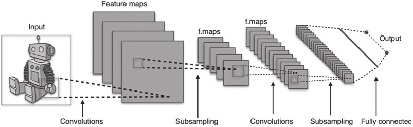
Figure 1.4 Architecture of a CNN (Creative Commons License).
become central to the operation of CV. This is due to DL’s many layers useful for identifying and modelling the different aspects of an image. In particular, convolutional neural networks (CNNs), a form of DL, involve a series of convolutions and max-pooling layers (see Figure 1.4) as its underlying architecture has been found to be very useful in image classification (AAIH, 2019; Erickson, 2019). CNNs are credited for reviving interest in neural networks in recent years. The way the CNNs work is by commencing with low-level features in the image and progress to higher-level features that represent the more complex components of the image. For example, the first layers will identify points, lines and edges, and the latter layers will combine these to identify the target class. An early example of CNN was AlexNet, an image classification model (AAIH, 2019). More recent versions are CNNs with specialised layers including ResNet, ResNeXt and regionbased CNN (Erickson, 2019).
CNNs are increasingly being applied in medical image interpretation (Erickson, 2019): for example, to classify chest X-rays that have malignant nodules and those that haven’t. Here, a set of labelled or annotated chest X-rays are used to train the neural networks to compute features that are reliable indicators of malignancy or lack. CNNs can be used for segmentation too where the class of interest is delineated from the remaining area of non-interest. However, CNNs are not restricted to analysing chest X-rays and have also been used to interpret CT, MRI, fundoscopy, histopathology and other images (Erickson, 2019; Reddy, 2018; Reddy, Fox, & Purohit, 2019).

