Learning and Cognition in Invertebrates
Invertebrates as Models
An emergent view in comparative psychology is that there is a remarkable similarity in learning and cognitive phenomena across species, perhaps reflecting a set of highly conserved cellular and molecular mechanisms underlying neural plasticity. The best way to assess generality is to study learning in species that are distantly related to the common laboratory models provided by mammals and birds. Chapter 4 described nonassociative learning in invertebrates, but the following sections center on studies of associative learning in species from several invertebrate phyla, including cnidarians, flatworms, segmented worms, and nematodes. Keep in mind that these species belong to different phyla and are as distantly related to each other as each of them is related to vertebrates.
One advantage of invertebrates is their simple nervous systems, which allow for the study of the cellular correlates of learning (see Chapter 4). These species are classified in phyla that were already differentiated in the early Cambrian, about 540 Ma (see Figures 2.2 and 2.5). Therefore, there has been a considerable amount of time for independent evolution. Moreover, the common ancestor of living animals with differentiated neural tissue (cnidarians and bilaterals) must have been a simple organism with a primordial neural system. Common learning mechanisms would imply deep homologies in synaptic plasticity, perhaps reflecting some fundamental constraints in cellular physiology and biochemistry.
The animal phyla depicted in Figure 7.3 can be grouped in three major categories. Sponges have no known neural tissue and thus are not useful in understanding the neural basis of learning and cognition. Radially symmetrical animals (cnidarians: sea anemones, coral, and jellyfish) have a diffuse neural network. Despite their simple organization, cnidar-ian nervous systems exhibit many properties also observed in neurons from other animals, including mammals. For example, chemical synapses are based on familiar neurotransmitters and neuropeptides (e.g., serotonin, acetylcholine, glutamate); both G protein-coupled and ionotropic synaptic receptors have been identified; some impulses are propagated in giant

Figure 7.3 The phyla of species used in learning and cognitive experiments described in Chapter 7 are represented in a phylogenetic tree depicting their evolutionary relationship and the emergence of some key traits. Sponges are shown as the outgroup; lacking neurons they have not been used in these types of experiments.
Source: All photos from Creative Commons, https://search.creativecommons.org: sponge by Velo Steve, license CC BY-SA 2.0; jellyfish by Fool4myCanon, license CC BY 2.0; flatworm by stupidhead, licensed CC BY-SA 2.0; nematod by NIAID, licensed CC BY 2.0; honeybee by tracie7779, license CC BY-SA 2.0; squid by Tracy Candish, licensed CC PDM 1.0; earthworm by Smabs Sputzer (1956—2017), licensed CC BY 2.0; frog by watts_photos, license CC BY 2.0.
axons; neurogenesis; and familiar ion channel receptors were described, including those for calcium and sodium, which play a key role in action potentials (Bosch et al., 2017). Not surprisingly given their nervous systems, cnidarians provide evidence of nonassociative (see Chapter 4) and associative learning. Classical conditioning was reported in the sea anemone Cribrina xantlwgrammica (Haralson, Groff, & Haralson, 1975). Animals were first habituated to Light, then exposed to Light/ Shock pairings, and finally to Light-only extinction trials. Control groups were given training with either Light-only, Shock-only, or “random” Light/Shock presentations. Two electrodes located at opposite sides of the anemone column recorded the contraction of the body and tentacles during the signal. Experimental subjects exhibited habituation during the initial Light-only trials, followed by a gradual increase in the amplitude of the contraction response during Light/Shock pairings, and by the extinction of this response during the final Light-only trials. CS/US pairings led to higher response levels than in any of the controls. This is an important finding given the simplicity of the diffuse nervous system of cnidarians, but, unfortunately, no analysis of this effect has been published.
The other phyla represented in Figure 7.3 are characterized by a bilateral body plan (see also Box 7.3). Even the simplest bilaterals, the flatworms, exhibit central and peripheral nervous systems. The planaria Dngesia dorotocephala and D. tigrina also exhibit evidence of classical conditioning (Baxter & Kimmel, 1963; Jacobson, Horowitz, & Fried, 1967). Animals exposed to Light/Shock pairings showed higher response levels than animals exposed to unpaired presentations of the Light and Shock, to backward Shock/Light pairings, and to simultaneous LightShock pairings. Thus, nonassociative effects are insufficient to explain
Learning and Cognition 245 these behavioral changes. There is conflicting evidence concerning the ability of planaria to form long-term memories; positive results were reported for memory of familiar places (Shomrat & Levin, 2013), but no evidence of retention for directional preferences modified by exposure to electric shock was reported (Abbott & Wong, 2008).
Box 7.3 Tardigrades
Tardigrades (commonly called “water bears”) are small animals (less than 1 nun long) that live in terrestrial or aquatic environments and have been classified in their own phylum: Tardigrada. More than a thousand species have been described, but the number has been increasing in recent years, so their taxonomy is being updated constantly (www.tardigrada.net/registei7taxonomy.htm). They are bilaterally organized and have a CNS that coordinates the movements of four pairs of legs. They deserve special attention because of a remarkable capacity. When exposed to unusually strong environmental conditions that would normally threaten the life of most animals (unusually low temperatures, extreme desiccation, maintained in a vacuum), tardigrades are able to shut down their metabolic processes and remain in a state of suspended animation called “tun” for days to months. When in a state of tun, their body dries out and the animal appears to be dead. However, when returned to normal environmental conditions, tardigrades revive and exhibit normal behavior. The mechanisms leading to a state of tun offer a unique opportunity to study metabolic aspects of learning and memory formation.
Very little is known about tardigrade learning. In one study (Zhou et al., 2019), individual tardigrades (Dactylobiotus dispar, see figure) maintained in water and fed with algae were isolated to determine their responses to various stimuli, including lights and electrical discharges. Their responses were observed under a light microscope and captured with a digital camera. In a key delay conditioning procedure, a Blue light (the signal) was presented for 20 s and followed by a 400-pA Shock (the reinforcer) lasting 2.5 s. The unconditioned response to the Shock was a shortening of the body.
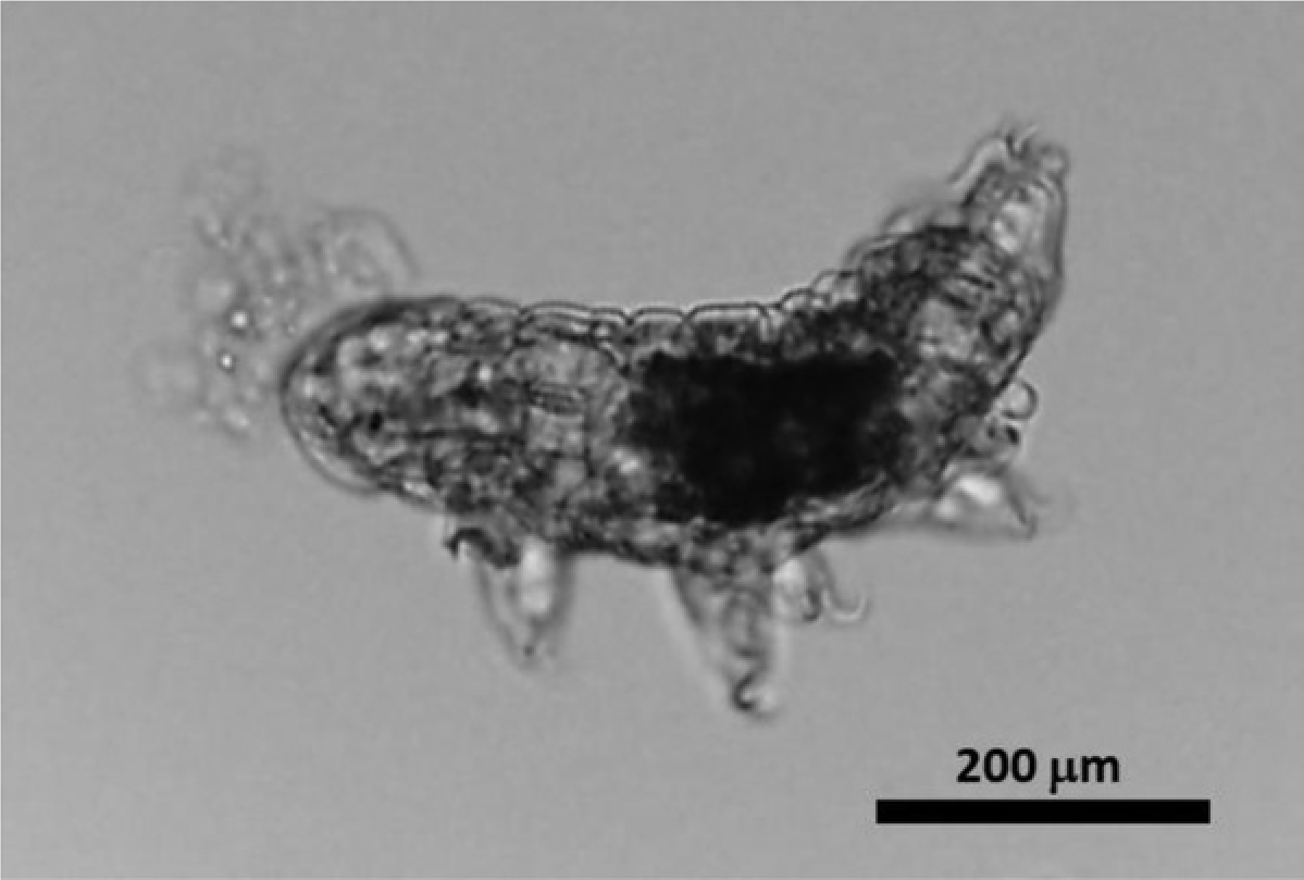
One hour after a single Blue^Shock pairing, animals exhibited a significant shortening of the body when exposed to the Blue light. Control conditions exposed only to the Shock, to a White light, to the Blue light without the Shock, to a backward Shock^Blue light pairing, or to random presentations of the Blue light and Shock displayed no changes in body length. Moreover, animals exposed to pairings of a Green or Red light with Shock also failed to exhibit a conditioned response. Whereas conditioning was detected when animals were tested one hour after training, animals tested four hours after training failed to exhibit a conditioned response, suggesting a rapid decay of this memory.
This procedure opens a door to study the effects of the interruption of metabolism on memory formation. Individual tardigrades could be trained, then exposed to conditions that induce tun, then reanimate, and finally determine what aspects of memory formation were affected by the metabolic arrest. (Photo by S. Pack, S. Blinova, and R. Ali, courtesy of Theodore Dumas.)
Experiments with earthworms (Lumbricus terrestris) show that pseudoconditioning plays a considerable role in aversive situations (Abramson & Buckbee, 1995). Worms respond to a neutral stimulus after US-only training, thus giving the impression of conditioning. Such pseudoconditioning can mimic conditioning in the absence of associative learning (see Box 6.2), reinforcing the importance of controls before establishing the capacity of animals to display associative learning. Another species of segmented worm, the leech (Hirudo medicin alts'), shows evidence of Pavlovian conditioning. When tactile stimulation (signal) was paired with electric shock (reinforcer) applied to the posterior end of the animal, the signal potentiated the shortening reflex, a defensive response that can be objectively measured (Sahley & Ready, 1988).
The nematode Caenorhabditis elegans allows behavioral studies to be combined with the physiological and genetic substrates of learning due to its striking simplicity (Ardiel & Rankin, 2010). The CNS of C. elegans contains just 302 neurons, all identified and with a known connectivity (WormAtlas et al., 2002-2018). In addition, the genome of C. elegans has been mapped, and available techniques, such as fluorescent imaging, permit in vivo genetic analysis of learning phenotypes. These experiments suggest that a simple neural circuit can be sufficient for classical conditioning. Consider the following demonstrations of associative effects, all with appropriate nonassociative controls:
- • Appetitive learning. A salt-based solution (signal) was paired with access to food (a layer of E. coli bacteria, the reinforcer). In a subsequent test, nematodes showed a preference for a location marked with the signal (Wen et al., 1997).
- • Aversive learning. Diacetyl odor (signal) was paired with exposure to an acetic acid solution (reinforcer). While nematodes were initially attracted to the diacetyl odor (tracking a spot placed in front of them), pairing experience reduced this tracking response below the level of unpaired controls exposed to both signal and reinforcer separated by 30-s intervals (Morrison, Wen, Runciman, & van der Kooy, 1999).
- • Contextual conditioning. Nematodes exposed to a mechanosensory stimulus (a stream of taps) responded with a reversed swimming response that habituates rapidly. When tested in the same context 1 h after training, the response was still habituated; however, when tested in a different context, the response showed recovery (Rankin, 2000).

Figure 7.4 Effects of two diff erent manipulations of the context on the retention of a habituated response in the nematode C. elegans. (A) Extensive exposure to the training context after training (extinction) leads to a loss of habituation. (B) Extensive preexposure to the training context before training (latent inhibition) leads to a failure of the context to control habituation.
Source: Rankin (2000); reproduced with permission from the American Psychological Association.
Furthermore, leaving the nematodes for 1 h in the training context without presenting the tapping stimulus led to a loss of habituation (contextual extinction), whereas leaving the nematodes for 1 h in the training context before habituation training retarded subsequent habituation (contextual latent inhibition; Figure 7.4).

