H-L Interactions in Positive Pressure Ventilation
When a patient is under positive pressure ventilation (PPV), the interaction between the lungs and the heart is elicited, and haemodynamic effects can be observed [1, 5].
The effects of airway pressure and volume can be addressed separately.
The inspiratory positive pressure is transmitted to the right atrium (RA) and vena cava and below the diaphragm to the abdominal district. A direct compression of the great veins in the cardiac fossa and of the RA wall by the lungs must also be taken into account. This process causes a reduction in the venous return to the right heart
[6] due to an increase in the difference between the transpulmonary pressure (TP) and filling pressure, which represents the gradient for the venous return. Such a reduction in the RV preload causes a reduction in the RV stroke volume and then a reduction in the cardiac output.
A specific aspect related to the effects of ventilatory pressure in the setting of mechanical ventilation in ARDS patients regards the decreased lung compliance that such a disease carries [7, 8]. In fact, a reduction in lung compliance leads to a reduced transmission of inspiratory positive pressure, with potentially fewer haemodynamic effects. However, if a patient with ARDS is kept at a constant tidal volume (TV), then his/her airway pressure will be increased regardless if compared to baseline healthy conditions; thus, both the intrathoracic pressure and the right heart pressure will increase and cause a reduction in the venous return. Therefore, despite a reduction in lung compliance in ARDS, the haemodynamic consequences of H-L interactions under mechanical positive pressure ventilation cannot be excluded.
The inspired volume mechanically delivered to the lungs has effects on both the alveolar and extra-alveolar capillaries. The alveolar capillaries are directly compressed, reducing the available pulmonary vascular bed; the extra-alveolar capillaries are simultaneously stretched with an opposite effect on their cross section. Taken
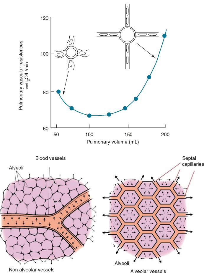
Fig. 16.5 Effect of pulmonary volumes on pulmonary vascular resistances. At low pulmonary volumes the effect is exerted on non-alveolar capillaries; high pulmonary volumes stretch the alveolar capillaries (Modified with permission from F. Guarracino “Ecocardiografia in Area critica” 2013 Elsevier, Milan)
all together, the net effect of the tidal volume on pulmonary circulation consists of an increase in the pulmonary vascular resistances (PVR) followed by an increase in the afterload to the RV (Fig. 16.5). Such an effect can add to the pressure-related preload effect, worsening the reduction in the RV stroke volume.
Again, a specific aspect regarding ARDS should be underlined: because the right ventricle acts as a “passive conduit” in normal conditions [5], ACP may be induced by adding the effects of PPV to the pulmonary vascular dysfunction occurring in ARDS, which leads to an abrupt increase in pulmonary artery pressure.
The H-L interaction under PPV leads to a fluctuating systolic arterial pressure waveform or stroke volume measure on the monitor screens whose variation represents a dynamic parameter to assess whether the patient is a fluid responder.
Interestingly, if a volume-related increase in the RV afterload predominates over the reduction in venous return, then the RV end-diastolic volume may increase with rising airway pressures, and an acute cor pulmonale (ACP) may develop [5]. This condition is not rare, being reported in up to 25% of patients with ARDS on mechanical ventilation [5]. If ACP occurs, then besides the RV dilatation and the reduction in cardiac output, other pathophysiological aspects characterising the acute ventricular interdependence deserve careful attention by the physician. In particular, it should be considered that in ACP, the RV undergoes a reduction of subendocardial flow, with myocardial ischaemia further contributing to impaired systolic function, hypotension and low systemic flow state and that the RV dilatation strongly affects the left ventricle (LV) filling. In fact, the pressure overload and dilated RV show a septal geometry ranging from flattened to leftward shifted, with the LV becoming acutely hypodiastolic [11].
In this scenario, tricuspid regurgitation may appear secondary to RV remodelling. IT adds prognostic information and affects the haemodynamic monitoring (cardiac output measured by PAC will no longer be reliable), impacting patient care.

