Pathogenesis
ARDS patients on MV often become colonized by exogenous pathogens acquired from healthcare personnel or, more commonly, by endogenous pathogens that colonize the gastrointestinal tract, oropharynx, tracheal tube, and proximal trachea. Several defense mechanisms prevent overwhelming colonization, i.e., cough, mucus clearance, and cellular and humoral immune responses. Yet, in ARDS patients, these defenses are impaired, because of the severe lung injury, comorbidities, and tracheal intubation. In the next paragraphs, several risk factors for the development of VAP will be described, with a specific focus on the specific risks and conditions that may occur in ARDS patients.
The Endotracheal Tube and Pulmonary Aspiration
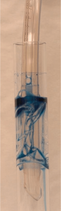
The primary mechanism in the pathogenesis of VAP is through pulmonary aspiration of colonized oropharyngeal secretions across the high-volume low-pressure (HVLP) endotracheal tube (ETT) cuff. The HVLP cuff was designed in the late 1970s to closely monitor the pressure exerted against the trachea [13]. Yet, when HVLP cuffs are inflated within the trachea, folds are formed, because the cuff outer diameter is larger than the tracheal internal diameter [14]. In vitro studies have demonstrated that cuff outer diameter, length, and positive end-expiratory pressure (PEEP) are the main determinants of cuff sealing efficacy. Indeed, the larger the cuff the more folds form on its surface, promoting leakage across the folds (Fig. 20.1).
Fig. 20.1 High -volume low-pressure cuff inflated within an artificial translucent tracheal model. The tracheal model internal diameter is 20 mm, whereas the cuff outer diameter is 28 mm. Upon inflation, folds are formed on the cuff surface. Methylene blue is poured above the cuff to highlight leakage of fluid across the channels formed by the folds
Pathogens may also grow on the internal surface of the ETT, forming a complex structure called biofilm [15]. ETT biofilm is composed by sessile bacteria within a self-produced exopolysaccharide matrix and respiratory secretions [16-18]. It is still not fully elucidated the role of ETT biofilm in the pathogenesis of VAP. Shah and collaborators [19] demonstrated a close association between intraluminal narrowing of the ETT by biofilm/secretions and length of stay on MV. Given that ARDS patients present longer periods of MV, it is reasonable to assume that a large amount of biofilm builds up and constitutes a persistent source of infection for recurrent episodes of VAP.

