FDM 3D Printing for Zygomatic Implant Placement Mock Surgery for Prosthodontic Dentistry: A Case Study
Dr. Priya Gupta, PG Student at VSPM Dental College, Nagpur, India, Dr. Saee Deshpande, VSPM Dental College, Nagpur, India, and Mr. Abhijeet Raut, Research Fellow, VNIT, Nagpur (MS), India, have done this case study of zyogomatic implant placement mock surgery for prosthodontic dentistry.
The quality of surgery depends on the anatomy of surgery. Three-dimensional (3D) anatomy visualization is very important for better understanding and a better outcome. Complex maxillofacial surgeries are usually time-consuming procedures due to the need for initial medical imaging for root complications, the determination of an appropriate course of treatment, pre-operative planning, before and after surgery. Pre-operative preparation is the most important and usually aims at executing an ideal procedure that minimizes the length of surgery and any possible problems that might occur in the future. For traditional dental procedures, the process begins by collecting the patient’s impression using a powder mold until it is moved to the dental grade of alginate powder coated. These topography data are generally sufficient for the majority of cases; however, they are less acceptable for intricate cases where an implantable device may be essential.
In addition, medical imaging data offer little insight, since patient representation is a collection of two-dimensional planar images. In such situations, 3D information on the internal bone tissue structure is necessary for optimal surgical planning in order to determine the precise geometry of the affected area of interest and to properly assess the size and orientation of the implant. More recently, 3D printing technology has shown tremendous potential to increase and streamline pre-operative surgery preparation and care.
Zygomatic Implant
In recent years, edentulous patients seeking fixed recovery have increased. Yet the condition is unacceptable when the maxilla is heavily resorbed and atrophic. A zygomatic implant can be a successful way to rehabilitate a badly resorbed maxilla. While zygomatic implants are responsive to the technique, they have predictable outcomes if performed correctly. The purpose of this case study was to modify the facets of the implant and to create a more patient-specific design. The idea of developing two abutment designs with an expanded base plate and modified abutment widths, lengths, and angles to suit the patient’s requirements has been discussed. Physicians and researchers took advantage of 3D printing technology and began a case study in 2018. The 3D printed prototype of the patient’s jaw was designed using CBCT data for ease of preparation and comprehension. A mock surgery was performed in the model as shown in the figure. The proper location of the implant and the expected result of the operation were evaluated. The final optimized design was validated by a finite element analysis (FEA) to analyze the force distributions that are likely to be observed after surgery. This process is vitally important to ensure that there are no structural flaws in the design, which could otherwise lead to a catastrophic failure. It was relevant in the present case, due to the high force exerted on the maxilla and mandible elements during eating and chewing and the ultimate goal was to ensure patient health and a good outcome of treatment (Figures 3.16 and 3.17).
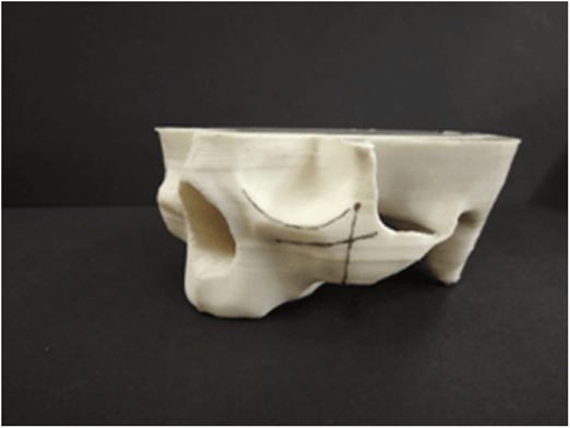
FIGURE 3.16 Shows the anatomical landmarks drawn in the model for the future position of zygomatic implants.

FIGURE 3.17 CAD model.
Conclusion
In this case study, Dr. Priya Gupta, Dr. Saee Deshpande, and Mr. Abhijeet Raut, along with the authors of the book, demonstrated the synergistic use of CT medical imaging, 3D CAD, and additive manufacturing in order to carry out the process of optimizing surgery planning and designing a personalized, patient-specific 3D model. And the 3D printing technology makes a complex process simpler for doctors. The use of medical imaging to establish a representative patient model has allowed the design of the zygomatic implant to be based on the specific anatomical characteristics of the patient. Using low-cost FDM 3D printing can also create medical models that allow mock surgery to be performed to test and optimize the design along with streamlining the procedure for actual surgery. Pre-surgical preparation has made the low-cost model possible and reduces the difficulty and time spent on the surgical innovation. This was also instrumental in achieving superior end esthetics and functionality. The findings of this case study provide a basis for future 3D printing of implantable devices. Such methodologies can result in lower costs for healthcare providers, thus increasing surgical effectiveness and enhancing the quality of treatment provided to the patient.
Exercises
- 1. Describe the concept of 3D Systems’ stereolithography apparatus (SLA).
- 2. Discuss Stratasys PolyJet with its advantages.
- 3. Explain fused deposition modeling from Stratasy.
- 4. Explain 3D Systems’ multi-jet printing system (MJP).
- 5. Discuss EnvisionTEC’s Perfactory.
- 6. Describe CMET’s solid object ultraviolet-laser printer.
- 7. Describe EnvisionTEC’s Bioplotter.
- 8. Discuss RegenHU’s 3D bioprinting.
- 9. Explain rapid freeze prototyping.
Multiple-Choice Questions
1. Stereolithography works on the concept of_a photosensitive resin
using a UV light layer-by-layer laser to create a 3D model.
- a) Solidifying
- b) Liquefying
- c) Modifying
- d) None of the Above Ans: (a)
- 2. Stereolithography was patented by 3D Systems in
- a) 1984
- b) 1986
- c) 1966
- d) 1989 Ans: (b)
- 3. Fused deposition modeling (FDM) was patented by Stratasys in
- a) 1984
- b) 1986
- c) 1966
- d) 1989 Ans: (d)
- 4. EnvisionTEC is the world’s leading provider of professional 3D printers and materials and was founded in
- a) 2001
- b) 2002
- c) 2010
- d) 2003 Ans: (b)
- 5. RegenHU 3DDiscovery is a 3D bioprinter developed by RegenHU, a manufacturer based in
- a) Germany
- b) Japan
- c) China
- d) Switzerland Ans: (d)
- 6. The aim of RegenFIU 3DDiscovery was to explore the potential of_
through a bioprinting approach.
- a) Bone engineering
- b) 3D tissue engineering
- c) Cell engineering
- d) None of the above Ans: (b)
- 7. Bioprinting is a precise deposit of_such as cells, proteins, bacteria, and bio-gels in 2D or 3D.
- a) Biocells
- b) Biochemicals
- c) Biomaterials
- d) All of the above Ans: (c)
- 8. Rapid freeze prototyping (RFP) is a relatively new solid freeform manufacturing process that builds a three-dimensional part according to the CAD model by depositing and_water droplets layer by layer.
- a) Freezing
- b) Solidifying
- c) Evaporating
- d) Liquefying Ans: (a)

