PRODUCTION OF BC COMPOSITES WITH SILVER NANOPARTICLES, BC/AGNPS
The dried BC films synthesized in the if. xylinus B-12068 culture had similar thickness (1.8 ± 0.2 nun) and density (0.15 ± 0.01 cm3), while their surface properties differed considerably, as determined by the water contact angle, which varied between 36° and 57°. The ultrastructure and size of fibrils in BC is a critical factor that determines the unique properties of BC films. SEM images of the microstructure of BC films show that BC films were layered nets of different densities composed of randomly oriented microfibrils (Figure 1.5). Whatever the drying method used, the average diameter of BC film micro fibrils was 110 nm, with the smallest and the largest diameters being 52 nm and 173 nm, respectively. However, in freeze-dried films, the fibrils were positioned more loosely, and the distance between them was 2.5-3.0 tunes greater than that in the films dried at room temperature, reaching about 1.6 pm.
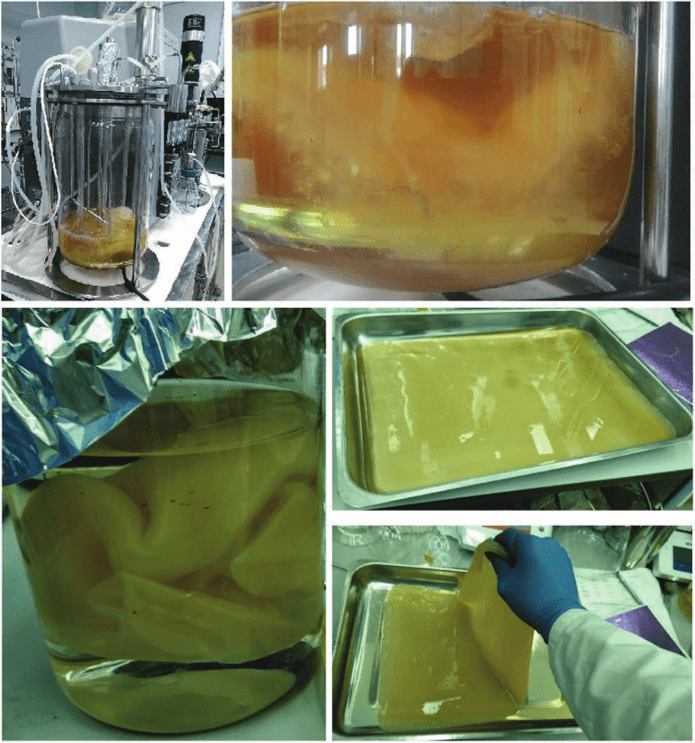
FIGURE 1.4 Scaling process of the bacterial cellulose synthesis in agitated culture in the fermenter and surface static culture.
The average crystallinity of the series of BC films was 60 ± 15%; the thermal decomposition temperature of the films was 260 ± 30 °C. The films had the following parameters of mechanical strength: Young’s modulus, 4.6-5.3 MPa; elongation at the break, 12-13%.
During the hydrothermal synthesis of silver nanoparticles, variations in AgN03 concentration in the reaction medium at 90 °C changed the number of the synthesized nanoparticles (Figure 1.6A) without considerably influencing their size (Figure 1.6B). For instance, the average sizes of Ag nanoparticles at AgN03 concentrations of 0.0001, 0.001, and 0.01 M were 13, 23, and 12 nm, respectively (Figure 1.7).

FIGURE 1.5 SEM images of initial BC films dried at room temperature (A) and freeze- dried ones (B). Scale bar =10 pm.
The increase in the number of silver nanoparticles in BC films is illustrated in Figures 1.7 and 1.8. The SEM images show that as the AgN03 concentration of the reaction medium was increased, the number of silver nanoparticles adhering to BC fibrils and between them increased (Figure 1.8). Ag nanoparticles showed different aggregation behaviors in freeze- dried BC films compared to those in the films dried at room temperature. On the films dried at room temperature, the size of Ag particles was 25-60 nm, the size of their aggregates was 85-350 nm, and the number of aggregates reached 15 per 1 pin2. On freeze-dried films, particle aggregates were larger, between 350 and 780 nm, and their number reached 19 per 1 pin2. The presence of silver in the BC/AgNPs was confirmed by the elemental analysis performed using scanning electron microscopy with a system of X-ray spectral analysis. The analysis showed that the average atomic number of silver particles in composite samples depended on the concentration of AgN03; as the AgN03 concentration in the reaction solution was increased from 0.0001 to 0.01 M, the silver content in the composites increased from 1.08 to 5.32.

FIGURE 1.6 Appearance of the reaction medium (A-l and A-2) in the process of obtaining composites of BC and silver nanoparticles at different temperatures and concentrations of AgNO,; size distribution of silver nanoparticles (В-1 and B-2).

FIGURE 1.7 Photos of composite films of BC/AgNPs obtained at different temperature conditions and AgNOs concentrations in the reaction solution.
One of the factors determining the biocompatibility of implants is the physicochemical reactivity of their surface. The main factors that influence surface interaction with blood and tissue cells and components of biological fluids are surface topography, microstructure, and adhesive properties. An indirect indicator of surface hydrophilicity is the liquid contact angle. The properties of BC composites are listed in Table 1.2. BC films are inherently quite hydrophilic, with the water contact angle below 50° (45.5° ± 17.6°). The surfaces of BC/AgNP composites tend to become less hydrophilic (more hydrophobic) as their silver content increases. The dispersive and polar components increase, too. Such changes reduce the fouling of the surfaces by proteins and cells, as described in a study by Jia et al. (2012). The authors of that study observed an increase in the water contact angle on BC/CuNPs composite films to 110 ± 0.4°—the value that was twice higher than that on the initial BC (50.1 ± 1.8°).
All samples of BC composites exhibited bactericidal activity against test microbial cultures—the most common representatives of nosocomial infection and pathogenic microflora of contaminated wounds. However, the

FIGURE 1.8 SEM images of the composites films of BC/AgNPs produced under different AgN03 concentrations in the medium: the fihns were dried at room temperature. Arrows denote aggregates of Ag nanoparticles. Scale bars = 200 and 300 pm.
levels of antibacterial activity of the composites were different (Table 1.3, Figure 1.9). ВС/antibiotic composites had a stronger inhibitory effect on the growth of pathogenic microorganisms.
TABLE 1.2 Surface Properties of BC/AgNP Composites
|
Samples |
Water Contact angles |
Dispersive Component Polar Component (mN/m) (mN/m) |
|
|
Pristine ВС BC/AgNPs |
45.5 ± 17.6 |
28 ± 9.24 |
17.5 ±8.37 |
|
0.0001 M BC/AgNPs |
50.3 ±4.61 |
42.6 ± 1.01 |
27.7 ±3.6 |
|
0.001 M BC AgNPs |
68.8 ± 1.76 |
46.2 ± 0.99 |
22.6 ±0.77 |
|
0.01 M BC/AgNPs |
69 ±2.71 |
44.9 ± 1.85 |
25.1 ±0.86 |
TABLE 1.3 Inhibition of Pathogenic Bacteria on the Solid Medium by BC/AgNP Composites
|
Samples |
Diameter of Inhibition Zones (nun) |
||
|
P. aeruginosa |
E. coli |
S. aureus |
|
|
Pristine BC |
- |
- |
- |
|
BC/AgNPs |
|||
|
0.0001 M |
12 ± 0.44 |
11 ±0.20 |
15 ±0.73 |
|
0.001 M |
13 ± 1.15 |
13 ±0.50 |
14 ±0.28 |
|
0.01 M |
14 ±2.11 |
14 ±0.61 |
15 ± 1.58 |
In experiments with BC/AgNPs, the largest zone of inhibition (15 ± 1.58 mm) was observed for S. aureus, at the highest concentration of AgNPs (0.01 M); the smallest zone of inliibition (11 ± 0.20 mm) was created by 0.0001 M ВС/AgNPs for E. coli. These differences are caused by the different susceptibility of bacteria to BC composites with Ag particles, their cell structure and physiology. Gram-positive bacteria are generally more susceptible to the bactericidal effect of AgNPs (Ruparelia et al., 2007). Similar data were reported by (Shao et al. (2016): in their study, at a concentration of BC-Ag of 0.01 M, the diameters of the zones of inliibition of E. coli and S. aureus growth were 11.7 ± 0.1 and 11.6 ± 0.1 mm, respectively. Feng et al. (2014) reported a study of BC composites with silver reduced using NaBH4, which created zones of inliibition of diameter about 16.1 mm for E. coli and 17.7 mm for S. aureus; the BC composites with silver reduced with sodium citrate created zones of inliibition with smaller diameters: 13.7 and 13.2 mm, respectively. The diameters of the zones of inhibition of B. subtilis and E. coli
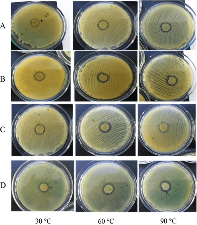
FIGURE 1.9 Inhibition of E. coli (A), K. pneumoniae (В), P. aeruginosa (C), and 5. aureus (D) with BC/AgNP composites obtained at different temperatures of the reaction medium.
by composites based on polydopamine magnetic BC and Ag were 20 mm and 17 mm, respectively (Sureshkumar et al., 2010). However, a study by Barud et al. (2011) showed that the composite membranes of 1 niol/L-1 BC/Ag/TEA created inhibition zones for P. aeruginosa ATCC-27853, E. coli ATCC 25922, and S. aureus ATCC 25923 reaching 20 mm. The differences between results may be caused by the different methods employed to produce composites, sizes of silver nanoparticles, and their concentrations in the BC films. It has been assumed that the lowest inhibitory concentration of Ag nanoparticles is about 0.05-0.1 mg/mL (Sureshkumar et al., 2010). In the present study, the strongest inhibition of pathogenic microflora by BC/AgNP composites was observed under the highest silver concentration in the reaction solution during composite production, when the silver content was 5.32 (or 0.024 mg/mL), that is, these results are consistent with the data reported by other authors.
The zones of inhibition by ВС/antibiotic composites were generally larger than those produced by BC/AgNP composites (Table 1.3). It is well known that antibiotics penetrate through the cell membrane and irreversibly bind to specific receptor proteins of the bacterial cell, thus effectively suppressing the synthesis of bacterial membranes.
As the experimental BC composites were investigated as possible candidates for wound dressings on skin defects and injuries, including those contaminated by pathogenic microflora, it was important to study the effect of silver and/or antibiotics impregnated into BC films on dermal cells. The BC composites were investigated for their potential cytotoxicity in fibroblast cell culture using 4',6-diamidino-2-phenylindole (DAPI) stain—a marker of nuclear DNA—and MTT assay, which determines the number of viable cells. The evaluation of the effect of BC composites on fibroblasts using DAPI staining was in good agreement with the results of the MTT assay (Figures 1.10 and 1.11). The results obtained in the present study are consistent with the published data, suggesting that BC/AgNPs produce a strong inhibitory effect on pathogenic and opportunistic pathogenic microflora, without inhibiting the growth of epidermal cells (Wu et al., 2014; Zhang et al., 2015).
No potential cytotoxicity was detected in all BC/AgNP composites in the NIH 3T3 mouse fibroblast cell culture, in contrast to the BC/antibiotic composites. These results suggest that BC composites constructed in the present study hold promise as dressings for managing wounds, including infected ones.

