Direct Electrochemical Oxidation of Blood
ABSTRACT
Protection of the human internal environment is an important aspect of the ecological problem. In a healthy human, this function is performed by the liver monooxigenase system that helps to remove hydrophobic toxic substances from the organism by their hydroxylating oxidation with molecular hydrogen catalyzed by special detoxicating ferment cytochrome P-450.
INTRODUCTION
The new area in efferent medicine—electrochemical oxidation of blood— is being successfully developed. Originally, the efficiency of direct current action on blood was found while working on fuel cells. Direct current was let to flow via various liquids, including blood. It was established that it destructively affects bacteria and their toxins.
In Russia, this method was developed in the works of the Academician Yu. M. Lopukhin. However, accidentally, the significant limitations of the method emerged. Under the electric field action, the blood corpuscles adsorbed, during the electrolysis in stagnant liquid in an electrolyzer, on the surface of electrodes and blocked them. The circuit current was interrupted. The attempt to recover the lost electrical conductivity by rigid mechanical actions upon the electrodes unblocked them and the electrolysis could continue as long as required, but, at the same tune, a number of other problems emerged, which prevented the development in this area. The investigations followed the way of electrolysis of sodium chloride aqueous solutions with further introduction of electrolysis products into the blood.
The process development received a boost after the elaboration of the method of direct electrochemical oxidation of blood by its electrolysis directly in the blood vessel. This technology significantly simplifies the method and the process instrumentation. The test on animals demonstrated its efficiency in various diseases. At the same tune, a new capability of the method, previously inaccessible, was discovered. As the electrolysis is conducted directly in the blood flow, it allows saturating the blood with elemental hydrogen. High hydrogen reducibility contributes to the reduction of nonequilibrium radicals in the course of cancer chemotherapy, facilitating the recovery process. This was the impulse to develop a variety of processes that allow selectively saturating blood either only with hypochlorite or only hydrogen, depending on the aim of the method application.
DIRECT ELECTROCHEMICAL OXIDATION OF BLOOD
Here, important is the issue of creating artificial systems modeling the liver detoxicating functions. This is conditioned by the fact that the existing methods of extracorporal detoxication (hemodialysis, hemosorption) are not efficient enough in removing hydrophobic toxins (Vasiliev et al., 1991).
Electrochemical methods can be used to clean blood and tissues from poisonous and ballast substances by their hydroxylating oxidation. The idea of modeling the liver detoxicating function with the help of electrochemical oxidation was formulated earlier (Yao and Wolfson, 1975). Despite the profound interest in the method of electrochemical oxidation, it did not find proper clinical application as, for the first time, only the principal capabilities of organism detoxication by direct electrooxidation of blood and other biological fluids were considered.
The systemic investigations were carried out to reveal the principal capabilities of electrooxidation of hydrophobic toxins in blood, lymphatic fluid, and plasma, to study kinetics and oxidation mechanism of typical toxins, to define the influence of electrochemical oxidation on different homeostasis indexes of intact animals, as well as to demonstrate how much effective is the electrochemical oxidation when used at the organism level (Vasiliev et al., 1991).
In this research, it was found out that electrooxidation of organic substances can proceed on different electrodes, but the majority of typical toxins are oxidized with a sufficient rate only on electrodes—catalysts of platinum group metals. A platinum electrode is the most suitable catalyst for modeling the hydroxylating function of cytochrome P-450 since practically all toxins are oxidized on it. The prelhninary research studies, conducted by different authors and referenced in this paper, are dedicated to the investigation of electrooxidation of various endogenic toxins on the platinum anode: bilirubin, cholesterol, urea, glucose, xenobiotics: methanol, ethanol, phenol, formaldehyde, and barbituric acid derivatives.
The device for electrochemical oxidation of blood in the form of an electrochemical cell with a volume of up to 150 mL equipped with input and output nozzles was tested. The cell is equipped with two electrodes, one of them is platinum. It is noted that under the same current load the increased electrode actual surface leads to a sharp increase in the blood trauma due to the increased number of blood corpuscles getting into the field of a double electric layer. Besides, during the blood contact with the electrodes, the development of blood clots is observed, the electrode surface gets rapidly covered with proteins and decomposed blood corpuscles, which block the electrode surface, and the electrooxidation process is stopped. The current does not pass through the cell. Two methods are proposed to respond to this phenomenon: the method of vibrating electrodes (Sergienko et al., 1984) and method of oxidation in the boiling bed (Vasiliev et al., 1986). The electrode vibration with a frequency of 22 Hz and an amplitude of 2 mm provides a normal passage of current through the cell. The method of oxidation in the boiling bed means that the process proceeds at the current high densities when its main part is spent on water electrolysis with oxygen and hydrogen evolution. The evolved gas bubbles form a boiling bed near the electrodes preventing the formation of a condensed film of proteins and blood corpuscles on the electrode surface. It is pointed out that both methods allow conducting the blood electrolysis for a long time. It should have generated some optimism in further works; however, in the course of extended systematic research studies, a broad range of the method applicability opportunities was found (Petrosyan, 1991); in addition, the method of direct electrochemical oxidation was considered unpromising at that time due to the complexity of practical implementation and low reliability of operation under clinical conditions.
Therefore, Petrosyan (1991) worked out and produced the device for electrochemical detoxication with indirect electrochemical oxidation of blood. The electrolysis of physiological saline is earned out in the electrochemical cell, and the product is introduced into the blood system with the help of a dropper tube. The method has shortcomings; thus, the volume of the solution introduced should not exceed 0.1 of the total blood volume at a low concentration of the final product, decreasing its efficiency.
Rudenok et al. (2004), for the first time, were able to solve the problem of blood corpuscles sticking onto the electrode surface and eliminate the effect of spontaneous stoppage of current flowing through the system by conducting the electrolysis inside the blood vessel without applying rigid mechanical actions upon metal electrodes. The mechanism of this phenomenon is still not entirely clear. Probably, the defining role is played here by “flow potential” preventing the blood corpuscles to nonreversibly adsorb as a dielectric barrier on the electrode surface. The method of direct electrochemical oxidation of blood envisages the electrolysis directly in the blood vessel based on bipolar polarization of the extended electrode. The process modeling in the electrochemical cell demonstrated that with the bipolar inclusion of the extended wire platinum electrode, the potential is distributed along its length, and at one of its ends the polarization potential corresponds to the oxidation process of chloride ion, and at the second one (cathode) the polarization potential corresponds to the potential of hydrogen reduction (Figure 12.1). Here, the detoxicating action is made by hypochlorite being synthesized but not by the electrical current in the blood (Rudenok and Marasinskaya, 2005; Rudenok et al., 2008).
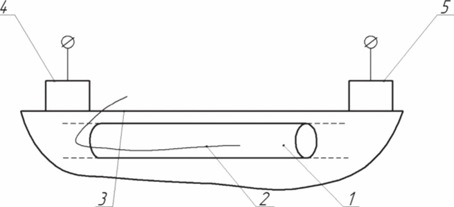
FIGURE 12.1 Scheme of direct electrochemical oxidation of blood. 1, blood vessel; 2, wire electrode; 3, skin covering; 4 and 5, applicator electrodes.
The hypochlorite action was positively tested on rabbits and calves with staphylococcal infection and pneumonia (Rudenok et al., 2004, 2011), and the method was introduced in one of the veterinarian clinics to cure skin conditions of dogs (Shabalina et al., 2006). More detailed investigations of the method using biochemical and other techniques were clinically made (Rudenok et al., 1999).
The experiments were conducted on 18 nonpedigree dogs of 1.5-2 years old, with the bodyweight from 20 up to 30 kg, selected by the principle of analogs. Three groups of six animals in each were formed: reference— clinically healthy, and the first and second test groups with the diagnosis of moist eczema on the back.
The reference animals underwent morphological and biochemical blood investigations, and the microelectrophoretic activity of their erythrocytes was determined with the help of the device “Cyto-Expert.” The data served as a reference. The dogs in the first test group underwent medical treatment following the scheme described below, without the direct electrochemical oxidation (DECO) of blood. The dogs in the second test group underwent medical treatment in combination with blood DECO. The eczemas of dogs were medically treated following the traditional techniques by parenteral administration of glucocorticoids (4 mg/animal), gamavit (following the instruction), and spot treatment with antiseptics (terramycin).
The animals in the second test group additionally underwent blood DECO with the help of the device for organism detoxication to treat endogenic and exogenic intoxication conditioned by the accumulation of various toxic substances. For this, the conductor of platinum wire was introduced into the forearm cavity of the lateral subcutaneous vein. Two bipolar electrodes connected with the direct current source were applied to the skin covering at end parts, and 3 mA current was imposed within 15 min. The procedure was repeated 10 times every second day.
Before the experiment and then after 1, 3, 7, 14, 21, and 28 days, all sick animals underwent clinical checkup, morphological and biochemical blood investigations, and the microelectrophoretic activity of their erythrocytes was determined with the help of the device “Cyto-Expert” for expressing the diagnostics of endotoxemia and microelectrophoresis of cells. The changes in the electrophoretic activity of erythrocytes in the process of eczema treatment by various methods allowed the authors to speak about the intoxication decrease and (or) its elimination.
The blood investigation of the dogs in the reference group demonstrated that the main morphological and biochemical indexes were within the physiological norm. The following blood changes of the dogs in both test groups were found before the treatment: decrease in the number of erythrocytes and hemoglobin, ESR increase, leukocytosis, eosinophilia, neutropenia, and lymphocytosis. The conducted investigations of the blood erythrocyte content when treating animals with eczema applying blood DECO demonstrated their number increase at all investigation stages. By the end of treatment, they exceeded the reference data by 2.78%. In the dogs in the first test group, even by the end of therapy, the index was by 8.33% lower in comparison with that of the clinically healthy ones. Hemoglobin in the blood of the dogs in the first and second test groups before the experiment was 104.0 ± 1.26 and 102.0 ±
1.11 g/L, respectively, and 125.0 ± 3.6 and 135.0 ± 4.0 g/1, respectively, after the experiment. In the dogs in the first test group, it did not reach the reference level by 6.72%, in comparison with the second test group. The data obtained indicate a favorable influence of this treatment method on the dynamics of erythrocytes and hemoglobin.
It was found that the amount of leucocytes in the blood of the dogs in the first and second groups starts decreasing during the treatment, but this process is more intensive in the dogs additionally treated with blood DECO. As can be seen from Table 12.1, the amounts of eosinophils, stab neutrophils, and lymphocytes decrease and approach the indexes of the reference group in 28 days after starting the treatment. This indicates the arresting of allergic and inflammatory reactions. Neutropenia and lymphocytosis, indicating the antigenic stimulation of the organism, disappear by the end of treatment. However, in the dogs in the first test group, the amount of lymphocytes decreases considerably, possibly being the reaction to the administration of glucocorticoids, and the immune suppression, resulting in organism weakening and emergence of a new pathology of these animals in the future, can develop. The dogs in the second test group, additionally treated with blood DECO, do not have such immune suppression that can be recommended as the way of immunity improvement.
The positive dynamics of the total protein in the animals of both test groups is observed. However, the most vivid changes are seen in the dogs treated with medicals and blood DECO. The increased level of protein exchange also indicates the improved immunity response from the annual organism as the reaction to blood DECO.
Aspartate aminotransferase (AST) and alanine aminotransferase (ALT) are important indexes in the evaluation of the medical effect of the above eczema treatment schemes. Before the treatment, the amount of AST in the animals in the first and second test groups exceeded the indexes of clinically healthy animals in 2.8 and 3.3 times, respectively. However, the AST content in the blood serum of the dogs in the first test group exceeded the reference data by 86.67%, even by the end of treatment, and in the dogs in the second test group, in which blood DECO was applied, by only 13.33%.
TABLE 12.1 Leukogram of the Dogs During Eczema Treatment by Various Methods
|
Indexes |
Animal Groups |
Before Treatment |
After 7 days |
After 14 days |
After 28 days |
|
1 |
2 |
3 |
6 |
7 |
9 |
|
Eosinophils, % |
Reference |
4.0 ±0.4* |
|||
|
1 test group |
7.5 ±0.47* |
5.8 ± 0.44* |
4.8 ±0.34* |
4.2 ±0.23* |
|
|
2 test group |
7.5 ±0.73* |
5.5 ±0.47* |
4.2 ± 0.44* |
4.0 ±0.40* |
|
|
Stab neutrophils, % |
Reference |
3.5 ±0.24* |
|||
|
1 test group |
5.2 ±0.18* |
4.3 ±0.37* |
4.0 ±0.40* |
3.7 ±0.37* |
|
|
2 test group |
5.3 ±0.23* |
4.8 ±0.18* |
4.3 ±0.37* |
3.3 ±0.24* |
|
|
Segmented neutrophils, % |
Reference |
62.7 ±0.83* |
|||
|
1 test group |
55.5 ±0.84* |
60.8 ±0.87* |
64.3 ± 0.67* |
69.3 ±0.97* |
|
|
2 test group |
55.7 ±0.73* |
58.2 ±0.91* |
60.5 ± 0.84“ |
62.8 ± 0.72“ |
|
|
Lymphocytes, % |
Reference |
28.1 ±0.5* |
|||
|
1 test group |
30.0 ±0.80* |
27.3 ±0.61* |
25.2 ±0.71" |
20.5 ± 0.4* |
|
|
2 test group |
29.8 ±0.72" |
29.3 ±0.83* |
28.8 ±0.66“ |
27.6 ±0.2* |
|
|
Monocytes, % |
Reference |
1.7 ±0.37**** |
|||
|
1 test group |
1.8 ±0.34*** |
1.8 ±0.34*** |
1.7 ±0.37**** |
2.3 ±0.37** |
|
|
2 test group |
1.7 ±0.37**** |
2.2 ± 0.34** |
2.2 ±0.34" |
2.3 ±0.37** |
Note: >'0.001, ">'0.002. ">'0.005, ***>'0.01.
Before the treatment, the ALT content in the blood of the dogs in the first and second groups differed from the indexes of clinically healthy annuals by
2.2 and 2.4 tunes, respectively. Despite the fast drop in the ALT amount in the animals in both test groups, the content of this ferment in the dogs, who underwent only the medical treatment, did not reach the level of the clinically healthy ones and differed by 1.4 tunes. In the annuals in the second test group, who were treated with blood DECO in combination with the basic traditional therapy, the ALT index reached the level of clinically healthy dogs.
Thus, blood DECO elicits the positive reliable dynamics of such laboratory indexes as AST and ALT, important during eczemas. The investigations carried out demonstrated that blood DECO inclusion into the treatment scheme during eczema treatment favorably influences the dynamics of alkaline phosphatase, total bilirubin, cholesterol, creatinine, and urea.
Since eczema is accompanied by intoxication, the authors applied the method of express diagnostics of endotoxemia based on microelectrophoresis with the help of the device “Cyto-Expert” (Table 12.2).
TABLE 12.2 Dynamics of Electrophoretic Activity of Erythrocytes in the Dogs During Eczema Treatment by Various Methods (P < 0.001)
|
Indexes |
Animal Groups |
Before Treatment |
After 7 days |
After 14 days |
After 28 days |
|
1 |
2 |
3 |
6 |
7 |
9 |
|
Oscillation amplitude of erythrocytes, men |
Reference |
13.69 ±0.71 |
|||
|
1 test group in % 1 to the reference |
5.49 ± 0.63 |
7.91 ±0.32 |
9.1 ±0.39 |
11.2 ± 0.61 |
|
|
40.1 |
57.8 |
66.5 |
80.5 |
||
|
2 test group in % to the reference |
5.58 ±0.53 |
8.99 ±0.41 |
10.7 ±0.38 |
13.4 ±0.52 |
|
|
40.8 |
65.7 |
78.2 |
98.0 |
||
|
Share of mobile erythrocytes, % |
Reference |
96.5 ±0.5 |
|||
|
1 test group |
59.9 ±0.6 |
71.8 ± 1.2 |
76.5 ± 0.6 |
83.7 ±0.5 |
|
|
2 test group |
59.1 ± 1.0 |
74.9 ± 1.0 |
81.2 ±0.7 |
96.0 ± 1.0 |
The investigations revealed the following. Before the treatment, the amplitude of erythrocyte oscillations in the animals in the first test group differed from the reference by 40.1% and in the animals in the second test group by 40.8%. By the end of treatment, this index maximally approached the data of the group of clinically healthy animals. The number of mobile erythrocytes in relation to their total number demonstrated the tendency to growth: from 59.9% ± 0.6 up to 83.7% ±0.5 and from 59.1 ± 1.0% up to 96.0 ± 1.0% in the dogs in the first and second test groups, respectively. However, this process was more intensive in the animals treated with DECO. Thus, blood DECO, in combination with traditional eczema treatment scheme, results in a reliable stable increase in the number of mobile erythrocytes in the animals of the second test group in a shorter period of time in comparison with the animals in the first test group treated only with medications that should prove the DECO detoxication effect.
Thus, blood DECO, following the above teclmique, activates the production of erythrocytes, white blood cells, increases blood hemoglobin, and also provides vivid biostimulating and detoxicating actions. Based on the aforesaid, the authors make the conclusion that blood DECO inclusion into the intensive therapy complex during dog eczema treatment improves metabolic processes, increases the activity of the organism protective factors, and contributes to faster arresting of inflammatory reaction and also the healing of the affected skin areas.
As indicated before, the problems of blood corpuscles sticking onto the electrode surface in the course of direct electrolysis in blood resulted in the necessity to develop the methods of indirect electrochemical electrolysis. As the process was conducted in a stagnant solution in a remote cell, it contributed to the permanent loss of the second important product of electrolysis-elemental hydrogen. In the process of long-term accumulation of hypochlorite in the solution, hydrogen completely volatilized from the liquid and was not considered by the experimentalists.
At the same time, it is known that the introduction of hydrogen into the blood decreases the necrologic effect in the presence of chemotherapy medications, not decreasing the therapeutic effect (Nakashima-Kamimura et al., 2009). Moreover, since in the process of electrolysis, the equivalent amount of hydrogen is evolved in the blood vessels simultaneously with hypochlorite, the authors attempted to use the electrolysis process in the blood vessel to accumulate hydrogen in the blood (Rudenok et al., 1999). This allowed coming close to the possibility of experimental detection of the efficiency of hydrogen obtained during the electrolysis as the deoxidant of nonequilibrium radicals being the reason of the side effects of chemotherapy in oncology. In this regard, it was necessaiy to expand the functional capabilities of the device due to the selective synthesis of either only hydrogen or only hypochlorite in the blood flow. For that puipose, the electrolysis technique was changed; the device scheme is shown in Figure 12.2.
The device consists of wire electrode 1, one part of which is submerged into blood vessel 2 and the second part is submerged into the attachable vessel 3 with the solution of electrolyte 4. Porous diaphragm 5 permeable for solution 4 is mounted in the bottom part of vessel 3. Additional electrode 6 is submerged into solution 4; the second additional electrode 7 is applied to the surface of skin covering 8 near the end of wire electrode 1 submerged into the blood. Electrodes 6 and 7 are connected to the direct current source (not indicated) via contacts 9, and vessel 3 is applied to skin covering 8 from the side opposite to the place, where electrode 7 is applied, with the possibility of electrical contact of solution 4 with the skin covering via porous diaphragm 5. When the electric current flows between electrodes 6 and 7 along the organism tissues, wire electrode 1, being on the way of the current lines, is polarized so that the part of wire electrode 1 submerged into the blood in blood vessel 2 is polarized to the potential with the sign opposite to the sign of electrode potential of electrode 7. The second part of wire electrode 1, being above the surface of skin covering 8 and submerged in the solution of electrolyte 4 inside attachable vessel 3, is polarized to the potential with the sign opposite to the sign of electrode potential of electrode 6.

FIGURE 12.2 Scheme of direct electrochemical oxidation of blood, which allows synthesizing only either hypochlorite or hydrogen in the blood flow (Patent no. 2566199).
Thus, wire electrode 1 is polarized by the bipolar scheme in such a way that its one end is polarized cathodically and the second one anodically. At the same tune, oxidation proceeds on the surface of one part of wire electrode 1 and reduction on the second one. Depending on the polarity of electrode 7, at the end of wire electrode 1, either chlorine ion in the blood plasma will be oxidized with hypochlorite ion formation or water molecules will be reduced with elemental hydrogen formation.
Accordingly, the opposite electrode processes will proceed in attachable vessel 3 on the surface of the part of electrode 1 submerged into solution 4. Consequently, the operation conditions of electrode 1 model the operation conditions of the bipolar electrode split by double diaphragms 5 and 8, excluding the penetration of the electrolysis products from one electrode space into another.
The literature review dedicated to the perspective area of blood electrochemical oxidation directly in the blood vessel, in particular, revealed a great interest of researchers in this direction. However, mostly, the problems were detected, the solution of which is still far to be clear. The experimental and ethical difficulties do not give a chance to rapidly complete all the tasks put. However, the promising perspectives can become an incentive to the in-depth study of this problem.

