Biosynthesis and Therapeutic Potential of Silver Nanoparticles
ABSTRACT
Prodigious properties of metal nanoparticles (NPs) result in their potential applications in various fields. The conventional methods involved in the synthesis of metal NPs are tedious, energy-consuming, and involve the use of toxic and hazardous chemicals. Thus, alternative methods involving the use of biological molecules such as enzymes, seed extracts, fruit extracts are the need of the hour. Such methods provide many advantages and are ecofriendly.
INTRODUCTION
Nanotechnology nowadays is impacting almost every aspect of human life due to its potential applications in the areas of electronics, material sciences, biomedical engineering, and medicine (Bhattacharyya et al., 2009). Nanoparticles (NPs) due to their large surface-to-volume ratio, surface functionalization, absorption in the UV-visible range, and controlled drug release are very important in human life. Generally, there are two types of approaches that are used for the synthesis of NPs, that is, top-down and bottom-up approaches. In the top-down approach, the bulk materials are reduced in the range of nanoscale. However, the bottom-up approach involves the joining of smaller atoms or molecules to grow to larger nanostructures.
Several synthetic methods, such as physical and chemical methods, are available in the literature for the synthesis of NPs (Bao et al., 2011). However, the chemical synthetic methods involve the use of hazardous or toxic chemicals and therefore such surface-functionalized NPs cannot be directly used for biomedical applications (Muklierjee et al., 2012).
Bioenthused synthesis of NPs provides many advantages over the traditional synthetic procedures as there is no need for harsh conditions such as high temperatures, high pressures, and toxic chemicals. However, it offers an economic, commercially viable, and ecofriendly synthetic route. Silver NPs find potential applications as antibacterial and antifungal agents in textile engineering and silver-based consumer products (Kumar et al., 2011; Zhang et al., 2008; Schrand et al., 2008).
The synthesis of metal NPs using plant extracts is a challenging aspect, but this method can be used for many therapeutic applications (Mittal et al., 2013; Mittal et al., 2012). Various types of plants have been used for the synthesis of metal NPs (Mittal et al., 2013; Li et al., 2007). Capping of the metal NPs with biological molecules offers the bioapplicability of such NPs in therapeutic activities (Iravani, 2011). The plant-mediated synthesis is quite cost-effective, simple, and provides safety (Shankar et al., 2004; Narayanan and Sakthivel, 2008; Huang et al., 2007; Li et al., 2007). The major issue is that when NPs are converted into the powder form, they agglomerate and thus lose their characteristic properties. To avoid such agglomeration, various types of capping agents are used. This chapter includes the synthesis of silver NPs using fruit extracts, germinating seeds, and seed extracts and the identification of the active components responsible for the reduction of metal precursors.
SYNTHESIS OF AG NPS USING FRUIT EXTRACT
One of the common methods for the synthesis of silver nanocolloids involves the reduction of an aqueous solution of silver nitrate (Sun and Xia, 2002). The synthesis of Ag NPs was characterized by the appearance of an absorption peak at 420 nm corresponding to the formation of various Ag NPs in the size range of 20-100 nm (Vigneshwaran et al., 2007). For the synthesis of Ag NPs, various parameters such as the pH, temperature, concentration of the fruit extract, concentration of the metal precursor, and reaction time were optimized (Mittal et al., 2014). Out of the above-referred parameters, it was observed that it is the pH of the reduction medium that plays a crucial role in the synthesis of Ag NPs.
Upon varying the pH, it was summarized that it is the neutral pH, that is, 7, where the increase in the yield of NPs was observed; thereafter, that is, at pH 8, it decreases and a further increase in the pH results in the agglomeration of Ag NPs (Table 3.1). Thus, pH 8 is the optimal pH for nanoparticle synthesis (Mittal et al., 2014) and it agrees very well with other reported literature works (Krishnaraj et al., 2012; Khalil et al., 2012).
TABLE 3.1 Summary of Various Optimized Parameters for the Synthesis of Silver Nanoparticles
|
Optimization of Various Parameters |
Corresponding Values |
|
Concentration of fruit extract |
50 pL/50 mL (from stock solution) |
|
Concentration of silver nitrate |
2 uiM |
|
pH of the medium |
8 |
|
Temperature of the medium |
35 °C |
|
Reaction time |
12 h |
The next parameter is the temperature, and it is observed that when the incubation temperature was increased from 25 °C to 35 °C, the absorption peak increased and thereafter it decreased. Thus, 35 °C was the optimal temperature required for the synthesis of Ag NPs. The incubation time, that is, the tune interval required, was observed to be 12 h. It was observed that after 24 h, due to the instability of Ag NPs, agglomeration occurs, and these results were in good agreement with the reported literature values (Khalil et al., 2012; Veerasamy et al., 2011). The stability of thus-synthesized Ag NPs was tested for 60 days with a 10-day interval using a UV-visible spectrophotometer. It was observed that the peak at 420 nm showed no alteration, thus confirming the stability of biosynthesized Ag NPs.
The size and zeta potential measurements of such particles indicated the Z-average value of 35 nm with a low polydispersity index (PDI) of 0.31 and a zeta potential value of-19.5. Thus, the negative zeta potential value clearly indicates that Ag NPs are mainly comprised of negatively charged groups and are responsible for the average stability of the NPs.
The transmission electron! microscopy (ТЕМ) micrograph of the synthesized NPs is obtained at 5-10 nm scales (Figure 3.1) and exhibits a spherical shape. The size range is 5-20 nm with a mean diameter of 12.5 ± 2.5 nm. Energy-dispersive X-ray spectroscopy (EDS) analysis also confirmed the presence of elemental silver in the NPs. X-ray diffraction (XRD) analysis confirmed the crystalline nature of the synthesized Ag NPs. It showed peaks at 20 values of 38.2°, 44.4°, 64.6°, and 77.5°, which are assigned to the [111], [200], [220], and [311] crystalline planes of the face-centered cubic (fee) crystalline structure of metallic Ag NPs, respectively (Narayanan and Sakthivel, 2011).
Fourier transform infrared (FT-IR) analysis shows strong absorption bands located at 1083, 1260, 1436, 1617, and 3373 cm-1. These absorption peaks were assigned to the strong stretching vibrations of C-N aromatic and aliphatic amines (1083 and 1260 cm-1), amide I band of proteins due to carbonyl stretching in proteins (1617 cm-1), and OH stretching in alcohols, flavonoids, and phenols (3373 cm-1), respectively. FT-IR analysis indicated the presence of flavonoid coating on the surface of NPs.
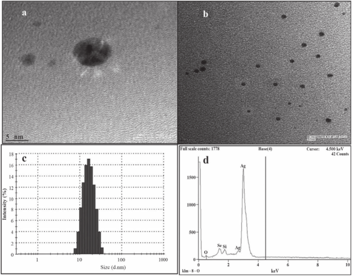
FIGURE 3.1 Enlarged ТЕМ image (a) 5 nm scale and (b) 10 nm scale, (c) Histogram of silver NP distribution, (d) EDS spectra of silver NPs synthesized by fruit extract [Reprinted with permission from Mittal et al. (2014). © Elsevier.]
To characterize the active compounds responsible for the reduction of silver ions, the solvent extraction method was used. It was concluded that flavanoids are mainly responsible for the synthesis of NPs. The following plausible mechanism (Figure 3.2) is proposed for the reduction of Ag~ ions to metallic silver through redox activity. The proposed mechanism agrees very well with the other proposed reports (Duran et al., 2005).
Antioxidant activity means the inhibition of the oxidation of the molecules by inhibiting the initiation step of the oxidative chain reactions that lead to the formation of the stable radicals that are quite nonreactive. Polyphenolic and flavanoid compounds present in plants possess strong antioxidant activity that protects the cells from various free radicals created as a result of oxidative stress.
The antioxidant activity of the synthesized Ag NPs was tested by various assays such as DPPH, ABTS, and MTT. The DPPH assay demonstrated an IC50 value of 22.19 pg/mL. while the MTT assay showed the antioxidant activity of 61% at the same concentration. The antioxidant property is due to the capping of Ag NPs by the flavanoids present in the plant extract.
Ag NPs were also tested for their cytotoxic effects on Dalton lymphoma (DL) cells using the MTT assay (Mittal et al., 2014). The Ag NPs showed a viability of 10% at 50 pg/rnL, whereas a viability of 46% was observed at 100 pg/rnL. Thus, they showed dose-dependent cytotoxicity. Such Ag NPs showed toxicity only on the tumor cells and not on the normal cells. Thus, they have the potential to be used as nanomedicine against various types of cancers. Thus, the applications of such ecofriendly NPs in bactericidal, wound healing, and electronic applications create the urge of large-scale synthesis of other metallic NPs.
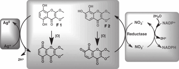
FIGURE 3.2 Prospective mechanism for silver NP biosynthesis (structures of isolated flavonoid molecules are shown) by the fruit extract of Syzygium cumini [Reprinted with permission from Mittal et al. (2014). © Elsevier.]

