BIOCHEMICAL VARIABILITY
The methodology followed for the following experiments on biochemical variability is as per Schaad (1992).
4.3.1 LACTOSE UTILIZATION
The carbon source (lactose) was filter-sterilized and mixed with autoclaved, cooled Dye’s medium along with 1.2% purified agar. The pH was adjusted to 7.2. Bacterial isolates were spot-inoculated with the replica plating method and incubated at 30 °C for 3, 7, and 14 days. The growth was compared with control, where the carbon source was not supplemented (Schaad, 1992).
4.3.2 STARCH HYDROLYSIS
The medium used for starch hydrolysis was sterilized by autoclaving and poured into sterilized Petri plates. These plates were inoculated and incubated at 30 °C for 7 days. The plates were flooded with Lugol’s iodine and allowed to act for a few minutes. The presence of starch hydrolysis was indicated by the presence of clear zones and vice versa. The hydrolyzed zone was measured for each isolate.
4.3.3 ACID PRODUCTION FROM SUCROSE, MALTOSE, AND DEXTROSE
Acid production by the isolate of X. axonopodis pv. pimicae was tested by using peptone water medium of Dye. In total, 10 mL of medium was dispensed in each test tube. This medium was sterilized in an autoclave for 15 min. To these tubes, filter-sterilized carbohydrates, viz., sucrose, maltose, and dextrose were added at 0.14% concentration. The tubes were inoculated with 0.1 mL of 24-h-old bacterial culture and incubated at room temperature for three days. A change in the color of the medium confirmed acid production (Figure 4.3).
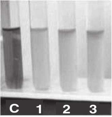
FIGURE 4.3 Acid production from sucrose, maltose, and dextrose (C, Control; 1, sucrose; 2, maltose; and 3, dextrose).
4.3.4 TESTING THE ANTIBACTERIAL ACTIVITY
After confirmation of X. axonopodis pv. ptmicae, the antibacterial activity of the plant extract was preliminarily screened by a well diffusion assay. Luria-Bertani (LB) agar plates were spread-plated with 20 pL of bacterial strain (1 x 10s cfu/mL). Wells of 6 mm diameter were made in the agar plates. Each plant extract was tested for antibacterial activity by adding 40 pL of extracts with different concentrations, viz., 50, 100, and 200 mg mL"1. The experiment was repeated thrice. The plates were incubated at 37 °C for 24 h. Subsequently, the plates were examined for the zone of inhibition (ZOI) and the diameter was measured (in nun) after subtracting the well diameter (Ahmad et ah, 1998). To determine the minimum inhibitory concentration (MIC), the required quantities of extracts were added to the LB broth of 4 mL to bring the initial concentration of 20 mg mL-1. In each test tube, 0.1 mL of standardized inoculum (lx 10s cfu/mL) was added. Two control tubes were maintained for each test batch, namely, extract control (tube containing plant extract and LB medium without inoculum) and organism control (tube containing LB medium and inoculum). The test tubes were incubated at 37 °C for 24 h. The lowest concentration (highest dilution) of plant extract that produced no visible growth (no turbidity) was recorded as MIC. The minimum bactericidal concentration (MBC) was assayed by subculturing test dilutions on a drug-free solid medium. The plates were incubated for 24 h at 37 °C. The lowest concentration of the antimicrobial at which no single colony observed after subculturing is regarded as MBC (Akinyemi et al., 2005).
RESULTS AND DISCUSSION
4.4.1 WELL DIFFUSION ASSAY
The preliminary screening of the selected three plant extracts against the X. axonopodis pv. ptmicae was done using a well diffusion method. The ZOI greater than 5 mm diameter is found to have significant activity against particular bacteria (Palombo and Semple, 2001). The aqueous extracts were sensitive to X. axonopodis pv.ptmicae tested at different concentrations (Table 4.3).
The extract of A. arabica was sensitive to X. axonopodis pv. ptmicae tested at different concentrations. The same extract was much effective at a low concentration of 50 mg mL"1, that is, 11 nun ZOI. P. juliflora extract arrested the growth of A. axonopodis pv. ptmicae (24 mm) at 200 mg mL'1.
The P juliflora extract shows ZOIs of about 21 mm and 19 mm at 100 mg mL'1 and 50 mg mL'1, respectively. Based on ZOIs, Satish et al. (1999) reported significant antibacterial activities of P juliflora and A. arabica against Xanthomonas campestris pathovars. The extracts of A. indicum exhibited the antibacterial activity of 11 mm ZOI at 200 mg mL'1.
TABLE 4.3 Antibacterial Activity of Plant Extracts Showing ZOI
|
Sr. No. |
Plant Species |
Cone, (mg ml"1) |
ZOI (mm) |
|
1 |
A. arabica |
50 |
11 |
|
100 |
15 |
||
|
200 |
16 |
||
|
2 |
P. juliflora |
50 |
19 |
|
100 |
21 |
||
|
200 |
24 |
||
|
3 |
A. indicum |
50 |
6 |
|
100 |
9 |
||
|
200 |
11 |
4.4.2 MINIMUM INHIBITORY AND BACTERICIDAL CONCENTRATIONS
The antibacterial activities of A. arabica and P juliflora extracts are found to be high against all of the bacteria. The maximum activity was recorded by P. juliflora (MIC = 1.03 mg mL"1 and MBC = 0.15 mg mL'1) and A. Arabica (MIC = 1.00372 mg mL'1 and MBC = 2.58 mg mL'1) against X. axonopodis pv. punicae. Raghavendra et al. (2006) reported the significant antibacterial activity of Acacia nilotica extracts against Xanthomonas pathovars and human pathogenic bacteria tested, while the lowest activity was recorded by A indicum (MIC = 0.619 mg mL'1 and MBC = 0.923 mg mL'1). These findings indicate that the extracts of P juliflora and A. arabica have the potential to be used in the management of plant diseases. Further phytochemical analysis is required to identify the active components of plant extracts showing antimicrobial activity (Table 4.4).
TABLE 4.4 MIC and MBC of Plant Extracts Against X. axonopodis pv. Punicae
|
Sr. No. |
Plant Species |
MIC (mg mL'1) |
MBC (mg mL'1) |
|
1 |
A. arabica |
1.00372 |
2.58 |
|
2 |
P. juliflora |
1.03 |
0.15 |
|
3 |
A. indicum |
0.619 |
0.923 |

