Examination of Alive Or Deceased Animal Gunshot Victims
Projectile injuries must be differentiated from puncture wounds, bite wounds, or lacerations. An animal with unexplained wounds should receive whole-body radiographs. Additional findings indicative of projectile injury may include pneumothorax, pneumomediastinum, cardiac tamponade, dyspnea, fractures, peritonitis, hemoabdomen, or hemothorax (Merck, 2013).
If possible, it is beneficial to have all the investigatory information and crime scene findings, specifically with regard to the type of firearm and ammunition, before the animal victim is examined. All confirmed gunshot wound victims must have whole-body radiographs. Consideration needs to be given that other injuries may be present such as those sustained from blunt force trauma. The skin may be reflected in deceased animals to define evidence of injury (Merck, 2013).
Examination of Alive or Deceased Animal Gunshot Victims
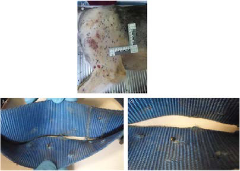
Figure 9.10 (a) Dog shot with shotgun (birdshot) from the left side, (b and c) Collar from dog shot with shotgun containing pellets from a shotgun round.
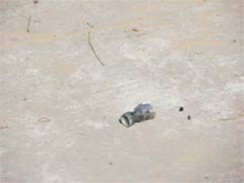
Figure 9.11 Wadding from a shotgun shell found near a dog's body.
In addition to a standard physical examination or necropsy procedures and reporting, the forensic veterinarian or pathologist must provide information regarding entry and exit wounds, projectile trajectory, and associated forms of tissue injury. The veterinarian must be aware of external evidence that needs to be collected as the animal is being examined (live animal) or prior to a postmortem examination to properly identify and retrieve ballistic evidence. Before initiating treatment ormanipulation of the carcass (if deceased) a plan should be created taking into account the best sequence of gathering evidence (Bradley-Siemens & Brower, 2016; Bradley-Siemens et al., 2018).
Before a gunshot case is encountered, a forensic veterinarian will need to communicate with local law enforcement agencies, crime scene analysts, or state crime laboratories, and be familiar with the types of evidence they can process and how evidence should be collected and submitted. These entities can offer expertise in evidence handling and collection and can direct trained personnel (ballistic experts) in obtaining and analyzing evidence such as gunshot residue and projectiles (Merck, 2013; Bradley-Siemens et al., 2018).
Photographs
Any wound surface or wound tract evidence should be collected only after photo documentation (Merck, 2013; Bradley-Siemens et al., 2018). The animal’s entire body will be photographed. Standard photographs will consist of front and back, left and right sides, and above (dorsal) and below (ventral). Each projectile wound should be photographed as it is found, then the fur shaved to identify wound characteristics or skin surface evidence and again photographed. All photographs will be taken with and without measurement (Merck, 2013).
Radiology
In any suspected gunshot injury, whole-body radiographs should be taken prior to a necropsy and at the earliest possible time frame, if the animal victim is alive. If a veterinarian will be actively participating in a forensic projectile casework, radiographic equipment must be available onsite or accessible through a working relationship with a facility that does. A minimum of two views with standard markers and identification needs to be obtained (Figure 9.12a,b). A radiograph will illustrate the shape and density traits of the projectile and the location of the projectile within the animal’s body (Figure 9.13a,b). Radiographs may define the pathway of the projectile within the body. Wound tracts may be defined based on the presence of gas, hemorrhage, bone, or metal opacities (Lockwood, 2013; Bradley-Siemens & Brower, 2016; Bradley-Siemens et al., 2018). The direction of fragments or beveling of bone in radiographs can assist with the direction determination of the projectile and define entry or exit wound identification (Figure 9.14). When a projectile strikes a bone, bone fragments will usually be found behind the damaged bone, showing the direction of the projectile (Bradley-Siemens & Brower, 2016).
Computed axial tomography (CAT) scans can be immensely beneficial in a forensic workup of either a deceased or live animal and, if available, is strongly advised by the author.
External Evidence Collection
Gunshot residue (GSR) is the result of the discharge of a weapon through gases and particles leaving the muzzle or through other openings in a firearm. GSR is the primer, powder, and/or projectile material and products of their combustion (DiMaio, 1999; Merck, 2013). GSR may be deposited around a projectile wound (as discussed in distance determination section) or on the hands of a shooter. This is a common type of external evidence found in projectile injuries.
Again, as protocols may differ between forensic laboratories, collection of external evidence should be discussed with these organizations prior to initiating this type of case. Depending if the animal victim is alive or deceased, tissue surrounding an entry wound may be excised. The skin and adjacent underlying soft tissues may be placed on a piece of cork or Styrofoam to maintain shape, size, and anatomical orientation for further analysis. A sample such as this can be analyzed using a scanning electron microscope and/or x-ray spectrometer to establish the quality and quantity of soot particles, information that can assist in range of discharge determination. There are several
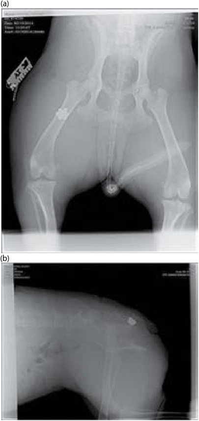
Figure 9.12 (a,b) V/D and lateral radiographs of gunshot wound to the thigh.
techniques used to collect evidence during an examination of projectile wounds. If powder particles associated with projectile wounds are found, they can be scraped into a Post-it note and placed in a sealed paper envelope. Entry and exit wounds can be shaved and the hair placed in a cellophane or waxed paper envelope. Clear tape can be utilized to obtain stippling from tissue and then placed on a glass slide. Areas of skin with contact wounds with soot or stippling can be taken at necropsy and either forwarded to a ballistic expert or photographed, sampled for residue, and fixed in formalin (DiMaio, 1999; Pavletic, 2010; Bradley-Siemens & Brower, 2016).
Although fired cartridge cases are usually observed at a crime scene surrounding or near an animal victim’s body, they may be accidentally collected when the animal’s remains are placed in a body bag and/or on the animal victim. A fired cartridge case can exhibit individual and class characteristics that can be used to identify a type of weapon or even a specific firearm. If a cartridge case is discovered on a killed or injured animal, it is important to save it as evidence (Haag & Haag, 2011) (Figure 9.15).
(a)

(b)
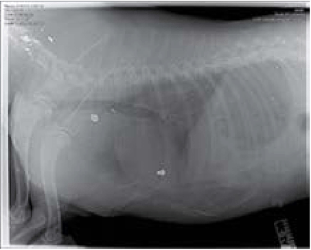
Figure 9.13 (a,b) Same dog shot multiple times with different types of ammunition, (a) 0.22 snake shot (pelleted rounds) to the face, (b) 0.22 rounds to the thoracic region.
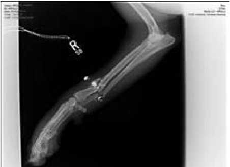
Figure 9.14 Gunshot wound to front of right leg, causing a radial-ulnar fracture and fragmentation of the projectile.
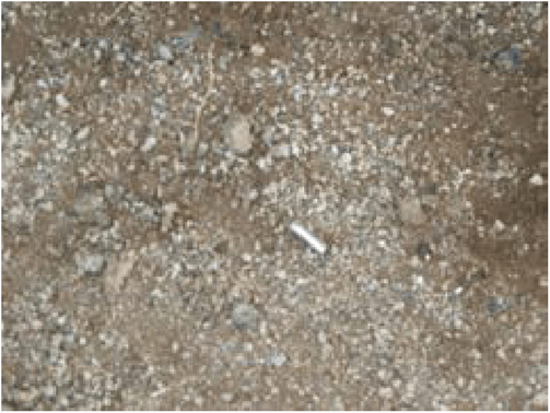
Figure 9.15 This is a cartridge casing found at the scene where a dog was shot, it is important that these be recovered for evidentiary purposes. Casings may be found in an animal's fur or in a body bag when remains are collected for necropsy. (Photo courtesy Phoenix Police Department.)
Gunpowder Analysis Techniques
Modern gunpowder contains nitrated cellulose. During the firing of a gun, particles of burnt, unburnt, and partially burnt gunpowder containing nitrite and nitrate compounds are emitted. Ballistic experts can utilize chemical analysis called a Modified Griess Test to detect organic nitrite residue on hair or skin (Jason, 2004; Haag & Haag, 2011; Bradley-Siemens & Brower, 2016). A ballistic expert can also use microscopy of fur or skin from entrance wounds to define particles of burnt or unburnt GSR (Jason, 2004; Haag & Haag, 2011; Bradley-Siemens & Brower, 2016). Infrared photography can be beneficial in demonstrating nitric acid presence in ammunition on the fur of an animal (Bradley-Siemens & Brower, 2016; Weiss et al., 2016). This can illustrate a residue pattern that can assist in determining a distance from which a projectile was fired. In addition, alternate light sources (ALS) and other tests such as sodium rhodizonate may be utilized in GSR detection and potential range determination (Weiss et al., 2016).
The ability to estimate firing distance to a target can be improved via various tests for GSR. The ability to conduct these types of tests will be on a case-by-case basis dependent on necessity and resources. It is advised that photography and visual enhancement be attempted prior to chemical tests that may alter or destroy a GSR pattern (Weiss et al., 2016).
Trajectory
The ability to define a projectile path or trajectory is important in the reconstruction of a crime scene or to corroborate or refute evidence provided by a witness or suspect. A trajectory can provide information about the location of a shooter, distance from an animal victim, and help determine the type of weapon and ammunition used.
To establish a trajectory, the veterinarian must first identify the entrance wound or wounds. Measurements of the length and width of a projectile entrance wound are obtained to establish the degree of angle of the wound. In addition, if GSR patterns exist these may further assist with directionality of the weapon discharge (DiMaio, 1999; Bradley-Siemens & Brower, 2016).
If the animal is deceased, trajectory rods help visualize and characterize a projectile’s trajectory in some cases. Rods must be straight, and may be made of fiberglass, plastic, or wood. The rods are meant to simulate the path of the bullet into and through an animal’s body. Rods must be placed with caution as to not create artificial tracts (Figure 9.16a,b). Once placed (animal in sternal
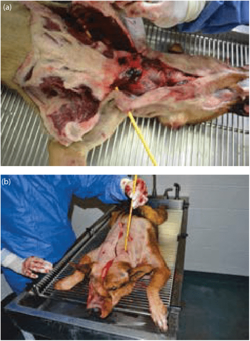
Figure 9.16 (a,b) Using a trajectory rod to assist in documenting projectile pathways through the animal.
recumbency), photographs should be taken from the front, back, both sides, bottom, and above the body. These photographs can assist in the development of theories based on investigation findings (Bradley-Siemens & Brower, 2016).

