Radiography and Imaging Signs of Neglect
Radiographs and additional imaging studies can benefit medicolegal investigations of animal neglect such as imaging evidence of failure to provide a minimum standard of veterinary care, imaging signs which support starvation, and imaging evidence supporting clinical findings of diminished body condition.
Subcutaneous fat and diminished intra-abdominal fat can be evaluated on radiographs. The measurement of subcutaneous fat surrounding the ventrodorsal thorax has been shown to correlate with the nine-point body condition score (BCS) in live animals (Linder et al., 2013).
The subcutaneous fat measurement is compared to the T4 vertebral body length and the T8 vertebral body width in a regression formula. With this method the body condition score could be radiographically determined when a body condition score was not performed or when the animal is no longer available.
Although radiographs may distinguish cortical and medullary bone and detect changes within the medullary cavity, MR is more sensitive for the identification of fat in the bone marrow. Serous atrophy of fat has been histologically identified in the bone marrow of emaciated animals and humans (Whiting et al., 2012). Increased fluid content of the bone marrow fat, consistent with serous atrophy, is identified on MR as a low signal on Tl-weighted images and a high bone marrow signal on short tau inversion recovery (STIR).
Radiology and Imaging Mimics of Abuse
Delayed presentation fractures, fractures in various stages of healing, bilateral fractures (Figure 18.13), and fractures inconsistent with the clinical history can be indicative of abuse in animals (Figure 18.14). No individual fracture location or fracture type is considered highly specific for nonaccidental injury in animals. Several disease processes can produce radiographic changes which could be mistaken for nonaccidental injury in animals. Endocrine disease, metabolic disease, and genetic disorders can produce multiple fractures in various stages of healing. Variations in ossification, normal growth plates, secondary centers of ossification, multipartite sesamoid bones, or skeletal anomalies must be distinguished from nonaccidental trauma. Periosteal new bone can occur secondary to disease processes unrelated to subperiosteal hemorrhage and trauma.
Increased radiolucency of bone is described as osteopenia and may be due to a variety of mechanisms (Figure 18.15). Osteoporosis, meaning increased porosity of bone associated with an imbalance in the normal homeostasis of bone and osteomalacia, indicating abnormal mineralization of bone matrix, is occasionally used interchangeably with osteopenia. Although most cases of

Figure 18.13 Ventrodorsal view of the thorax in a cat with multiple bilateral rib fractures and hemothorax. Mild periosteal new bone surrounds the rib fractures, indicating healing.
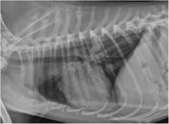
Figure 18.14 Lateral thoracic view of a rescue dog with unknown history. Multiple healing spinous process fractures and a pulmonary hematoma are present.
osteopenia are due to osteoporosis, causes of decreased bone opacity cannot be determined from radiographic evaluation and in such cases the term osteopenia would be preferable. Osteopenia is recognized on radiographs as a decreased medullary opacity in bone and cortical thinning. The diminished visualization of number and size of trabeculae in the ends of the long bones may be the earliest finding of decreased bone opacity. The identification of cortical thinning in osteopenia

Figure 18.15 A lateral projection of the antebrachium in a dog with a chronic malaligned nonunion fracture. There is generalized osteopenia in the manus consistent with disuse.
generally occurs later than the medullary changes. In addition, radiographs are not very sensitive for identification of bone mineral loss, and at least 30% bone mass loss is required before radiographic detection is possible. Without comparison to normal bone and without densitometry, generalized osteopenia is difficult to radiographically confirm and can be an artifact of overexposure. CT is superior to radiographs for detection of bone mineral density. Hounsfield unit values in regions of interest (ROI) are converted to bone mineral opacity by comparison to a bone density phantom through linear regression analysis. Quantitative CT has been validated in poultry and dogs to determine bone mineral density (Korver et al., 2004).
Endocrine disease, metabolic disease, reproductive status, and aging may contribute to an increased porosity of bone, predisposing an animal to fracture. Without bone densitometry, bone loss can be present prior to the radiographic identification of osteopenia. Endocrine diseases such hyperadrenocorticism, hypothyroidism, hyperthyroidism, and primary hyperparathyroidism can be associated with osteopenia. The radiographic identification of osteopenia on radiographs of patients with hyperadrenocorticism has been questioned; however, bone loss secondary to exogenous steroids is known to occur in animals with Cushing disease and can also be expected in any animal on chronic steroid therapy. Imbalances of parathyroid hormone, thyroid hormone, and estrogen may also result in osteoporosis.
Any disease process or diet that results in a change in circulating PTH, calcium, phosphorous, or vitamin D can result in fibrous osteodystrophy, which is increased osteoclastic or resorptive activity of bone replaced by fibrous connective tissue. Radiographically, the medullary cavity will enlarge and the cortices will appear thinner. The earliest changes are seen in the skull, where increased lucency is present in the bone surrounding numerous teeth and moth-eaten lysis can be visible in the calvarium. Joint subluxation may be apparent on imaging studies. As the disease progresses, thin cortices and medullary lucency will be observed in the long bones and multiple bone fractures may be recognized. Because of the softening of the bones, the fractures are often described as folding fractures. In young animals with fibrous osteodystrophy, the physes remain normal.
In immature animals presenting with multiple fractures without history of trauma and without evidence of nutritional or renal hyperparathyroidism or endocrine disease, an underlying genetic disease may need to be ruled out (Figure 18.16a,b). Osteogenesis imperfecta is a hereditary disorder resulting from defects in the genes responsible for the development of collagen. Animals and humans with this disease can present with multiple pathologic fractures, including fractures of the teeth. An overall diminished bone opacity and thin cortices may be appreciated on radiographs. Osteogenesis imperfecta has been reported in several dog breeds, including the dachshund, beagle, golden retriever, poodle, Bedlington terrier, collie, and Norwegian elkhound, and in cats. Reports in other animals, some of which are used as research models for osteogenesis imperfecta in humans, include mice, zebra fish, sheep, tigers, and cows. Genetic testing is available in certain dog breeds to determine carrier or affected status of osteogenesis imperfecta. Fibroblast cultures from a skin biopsy can be used for confirmation in other animals.
A silicate-associated osteoporosis or equine bone fragility disorder has been identified in horses in California. Affected horses without a history of trauma can present with acute fractures and fractures in various stages of healing and lysis of multiple bones.
Rickets is a metabolic disease which can occur in immature and mature animals. The radiographic findings may be similar to the findings of osteoporosis; however, the disease is due to a failure of the bone matrix to mineralize or osteomalacia, rather than an increased bone resorption. Rickets is primarily caused by a dietary deficiency of vitamin D or phosphorus. Inherited forms of rickets have also been reported in cats, dogs, pigs, and sheep. Llamas and alpacas outside of the Andes are prone to develop rickets, most likely due to a dietary deficiency of vitamin D and decreased exposure to sunlight during the winter months. Radiographic signs of rickets may include widened physes, flared metaphyses, thin cortices, decreased bone opacity, and pathologic fractures. Changes

Figure 18.16 Ventrodorsal (a) and lateral (b) pelvic views of a kitten with suspected osteogenesis imperfecta after ruling out metabolic disease. Note the folding fracture in the left femur and the right femoral neck fracture.
are most pronounced in the tibiae, radii, metacarpal bones, and metatarsal bones. Costochondral enlargements are also described and should be differentiated from normal costochondral remodeling.
When evaluating animals with multiple fractures and little evidence of trauma, three additional conditions or mimics of nonaccidental injury are considered: (1) small-breed dogs presenting with single or bilateral antebrachial fractures and a history of a jumping down or minor falling down event; (2) multiple dog breeds, particularly cocker spaniels, Brittany spaniels, Labrador retrievers, and Rottweilers, presenting with single or bilateral elbow fractures after minor trauma; and (3) young cats presenting with bilateral femoral fractures and no history of a traumatic event.
Distal antebrachial fractures are reported to be the third most common fracture in dogs, and small-breed dogs have a tendency to sustain distal antebrachial fractures with minor falls or jumping. These fractures, usually transverse, tend to be located in the distal one-third of the antebrachial diaphysis and can be confirmed on physical examination and radiographs. Bone density and geometric properties such as cross-sectional area, cortical thickness, and properties of shape that determine deflection are potential reasons for the propensity of small-breed dogs to sustain antebrachial fractures with minimal bending or torsion (Brianza, Delise, Ferraris, Amelio, & Botti 2006).
The canine humeral condyle develops from three ossification centers: one for the medial humeral condyle, one for the lateral humeral condyle, and one smaller center for the medial humeral epicondyle. The physis between the medial and lateral condyles will fuse by 12 weeks of age. In some spaniel breeds, Labrador retrievers, and Rottweilers, a predisposition to fracture at the physis has been observed either due to incomplete or delayed closure or a tendency for stress fracture at this site. Radiographs are sufficient to confirm a condylar fracture and may detect incomplete ossification; however, CT is more sensitive for evaluation of the physis and any adjacent sclerosis.
Cats greater than 1 year of age may present with bilateral femoral capital physeal fractures and no history of trauma. Although the femoral head physes typically close between 6 to 9 months in the cat, spontaneous unilateral or bilateral femoral head physeal fractures may be observed in cats greater than 1 year of age. The femoral head physeal fractures are diagnosed by routine pelvic radiographs, including a lateral and extended-leg or frog-leg ventrodorsal views.

