Congenital Deformities/Abnormalities from Inbreeding
Inbreeding of animals can result in a higher than normal appearance of congenital abnormalities, as it can concentrate the occurrence of recessive genes and the defects they cause. Reputable breeders carefully examine the pedigree of the potential breeding mates to avoid or minimize such defects. Breeding in hoarding situations, puppy mills, and disreputable rescues/shelters is usually indiscriminate. Often the dominant male will breed multiple generations and thereby concentrate the gene pool. In one hoarding case, in which the animal owner was convicted of animal cruelty, two litters of mixed-breed puppies were diagnosed with cerebellar abiotrophy (author’s professional experience). This inherited disorder is a fairly rare degenerative cerebellar condition seen in breeds such as Kerry Blue terriers, Australian Kelpies, and rough-coated Collies (Chrisman, 1991; De LaHunta, 1977; Kahn, 2010).
Matted Coats
Some breeds of animals have naturally long and/or curly coats that require regular clipping and grooming. In neglect cases, some animals may present with their hair coat as one solid mat. The matting may extend from the top of their heads, across their face obscuring the animal’s vision, and/or down to their paws (Figure 16.7). In these cases, consideration should be given to collecting the following data: photographs of initial presentation; weight of the animal prior to clipping; photographs during the clipping process; photographs of the animal after the fur has been removed to include all lesions; photographs of the paws, as often this severe matting can cause secondary pododermatitis (skin infection of the paw) lesions; weight of the removed hair; photographs through the holding period during the course of the case to document the appropriate routine grooming of the animal; and full documentation of all routine grooming required during the course of the
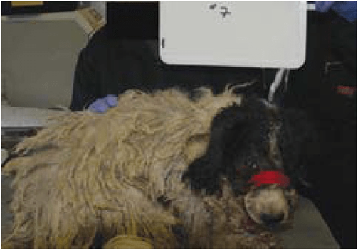
Figure 16.7 Dog with matted hair coat.
case and its associated costs. It may also be advisable to vacuum-pack the hair mat as evidence and document it photographically as well.
External Parasite Infestations
In neglect cases, either single-animal or large-scale cases, external parasite infestations are common. Full documentation of the extent of the infestation(s) is necessary for each case and for each animal involved in the case. The author has had the experience of having a large number of fleas jump on the personal protection equipment (PPE) to the point of discoloring the PPE upon entry into a hoarded residence. Since these parasites are easily visible on a white Tyvek suit, photographic documentation of this occurrence is recommended.
Tick infestations are fairly common, depending on the geographic location of a scene and the season. Whenever possible, documentation of the number of ticks on the animal is beneficial. In some cases, the infestations are such that actual counts are impractical. In this case, estimates with photographic documentation can be used (Figure 16.8a-e). If the notation of "TNTC” (too numerous to count) is used, then an explanation of the threshold for use of that designation is recommended. For example: “TNTC” is used whenever the number of parasites exceeds 1000. In the event of a tick infestation, ancillary testing for tick-borne diseases such as Anaplasmosis, Rocky Mountain Spotted Fever, and Lyme disease is warranted. Presence of one or more of these diseases is further evidence of the harm done to the animal by the neglect of the caretaker as tick infestations, and subsequent disease transmissions, are preventable with routine use of over-the-counter (OTC) medications.
Lice can be found in situations of crowding and/or poor husbandry. Avians with lice may show poor feather conditions, plucked areas, or areas of broken feathers. Mammals may show areas of alopecia (missing hair), excoriations (scratches), and/or raw spots (Kahn, 2010). Documentation of the overall condition of the animals and their environment, photographs of the parasites, identification of the parasites, documentation of the treatment of the animals and the subsequent improvement of the animals will substantiate the neglect and harm caused. As lice and other external parasites can be found in cages, kennels, and bedding, if any of these items are removed from the scene, precautions should be taken and the items sanitized before they can contaminate another area. The sanitation measures and justifications should also be documented in writing and with photographs.
In hoarding situations and disreputable dog/cat/rabbit rescues/shelters, ear mite infestations can be rampant and severe. Due to failure to treat this condition, the ears may develop polyps, scarring,
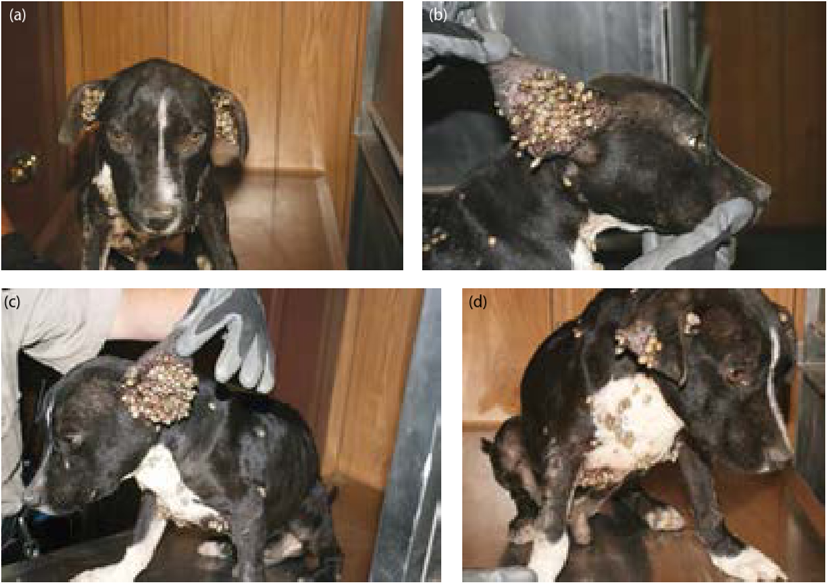
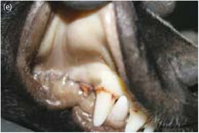
Figure 16.8 (a) Tick infestation, front view, (b) Tick infestation, right view, (c) Tick infestation, left view, (d) Tick infestation, chest view, (e) Pale mucus membranes of dog with tick infestation.
pinna (ear flap) alopecia (hair loss), and ulcerated, bleeding lesions within and on the ears. Cats with severe ear mite infestations are extremely resistant to handling or treatment of the ears due to the severe pain and discomfort from this condition. Quite often these cats require heavy sedation or anesthesia in order to clean and treat the ears. Ear mites can also be seen in dogs, especially puppies housed in unsanitary conditions (Kahn, 2010; Medleau & Hnilica, 2001).
Mange, both sarcoptic (scabies) and demodectic (demodex), is caused by external skin mites and are often seen in neglect cases. The lesions in scabies may initially present as alopecia around the pinnae margins and may have progressed to complete alopecia of the entire body with secondary changes such as lichénification (wrinkling) of the skin, hyperpigmentation (darkening of the skin), and ulcerated sores. Scabies causes intense pruritus (itchiness) and the dogs often scratch themselves endlessly. The scabies mite is zoonotic and has been known to cause similar lesions in humans. Therefore, care should be taken to mitigate transmission to humans and other animals when handling and housing these animals. Demodex is most commonly found in young and/ or immunocompromised animals. Malnutrition can cause immune incompetency which could predispose and/or worsen the condition in a susceptible animal. Demodex mange can result in severe secondary bacterial infections, and in some cases progress to septicemia. Depending on the animal’s capability to become immune-competent, the treatment for demodex may be prolonged and/or lifelong. Demodex mange is not zoonotic (Kahn, 2010; Medleau & Hnilica, 2001).
Birds housed in crowded and/or unsanitary conditions can also be infested with several different species of mites, some of which will feed on humans as well (Kahn, 2010).

