Deceased Animal Fire Victim
It must be determined if the animal was alive or dead prior to the fire or if there is evidence of antemortem injury. There may or may not be evidence of external injury. Those bodies with minimal or no external injury may have succumbed to smoke inhalation. Some bodies may have external signs of searing damage causing the skin to be light brown and have a stiff leathery appearance (DiMaio & DiMaio, 2001) (Figure 12.8a-b).
The animal’s body may be charred and swollen from heat. Heat accumulation in the thorax can cause blood to infiltrate the alveoli, airway, mouth, and nostrils, simulating an antemortem injury.
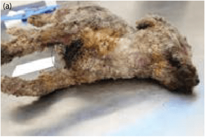

Figures 12.8 (a,b) Dog placed in BBQ; 40% of dog's exterior surface shows burning, scorching, and singeing of the fur and integument. (Photo courtesy Dr. J.D. Struthers.)
The presence of blood in the chest pleura (inner lining) can indicate antemortem trauma (Stern et al., 2014). The lung tissue may present with accelerant residue if the animal inhaled any of the vapors antemortem (Merck & Miller, 2013). In situations of a severely burned body, the skin may shrink, causing it to split. In other instances, with skin completely burnt away the underlying muscle may be exposed and can be ruptured due to the heat. Muscle will split parallel to the muscle fibers due to the heat traversing. Splits across the muscle may be due to trauma prior to death (Spitz, 2006; Merck & Miller, 2013). There may be areas of unaltered skin where the body was lying on a flat surface. The internal body wall may be removed from burning, exposing viscera which can be seared or charred (DiMaio & DiMaio, 2001; Merck & Miller, 2013).
It is very difficult to determine antemortem versus postmortem burns on gross examination. There may be microscopic evidence of vital response (inflammation); however, the lack of response is not indicative of postmortem injury. Thrombosis from heat in the dermal vessels may impede inflammatory cells from reaching a burn area (DiMaio & DiMaio, 2001; Merck & Miller, 2013).
Two dogs were found deceased in a house fire with a postmortem interval of approximately 24 hours. One of the two dogs had severe burns on over 50% of its body (majority on the dorsum and right body wall) with severe charring and splitting of the skin. No singe marks or soot deposit were observed on the eyelids. Internal examination of both dogs demonstrated deposition of soot (black pigmented material) mixed with mucous within the pharynx, larynx, trachea, and primary bronchi. The oral mucous membranes and musculature were red. The microscopic findings were limited to the respiratory system of both dogs. The was soot overlying the ciliated epithelium of the trachea and bronchi and the epithelium of the bronchioles. There was only minimal soot within the alveolar spaces (Stern et al., 2014).
In the deceased animal, blood collection and the determination of CO levels along with evidence of inspiratory activity (see above) are the best indicators an animal was alive before and during a fire. Whole blood can be obtained from the aorta, heart, or caudal vena cava. In conversations with Dr. Stern (Stern et al. 2014), blood samples for CO were collected in a green top heparin tube. These samples will need to be sent to a human laboratory or hospital for analysis. These samples should be obtained as soon as possible and sent for analysis (Stern et al., 2014).
In a fire, blood and marrow can be exuded from the skull and amass between the bone and dura mater, simulating a traumatic epidural hematoma present in the frontal, parietal, and temporal regions with possible involvement of the occipital region. The amassed material is a clot-like chocolate-brown color with a crumbly or honeycomb appearance (DiMaio & DiMaio, 2001; Merck & Miller, 2013). One indicator that an epidural hematoma is not antemortem is the presence of a skull fracture due to heat changes; this is absent antemortem. According to Spitz, subdural hemorrhage is an indicator of an antemortem injury (Spitz, 2006; Merck & Miller, 2013).
A burnt body may present with flexion of the limbs, causing a pugilistic posture. This is due to coagulation of the muscle tissue and contraction of muscle fibers from generation of heat when the body is burnt. The resulting muscle shrinkage may cause bone to fracture. The use of computed tomography can assist with differentiation of traumatic versus thermal changes (Merck & Miller, 2013).
Entomology
There may be evidence of live or deceased insects or larvae on burnt animal victims both internally and externally. After some time, insects present on the body can be the same first-wave insects as those found on fresh remains (Merck & Miller, 2013). These can assist with the postmortem interval. This can help assess when the fire occurred. Deceased entomology evidence (larvae) can be found internally, specifically in the skull or body cavities. This can indicate the animal was deceased before the fire (Merck & Miller, 2013).

