Bone Structure and Growth
Bone is made up of protein (collagen) and mineral (hydroxyapatite) portions. Collagen, the organic component, constitutes approximately 30% of a bone’s content, while the mineral portion makes up approximately 65% of a bone’s content, and the remaining 5% is water. Collagen gives bone the toughness and elasticity needed to flex and endure tension, while hydroxyapatite gives it the rigidity and hardness needed for stability (Romer & Parsons, 1977). Keratin is the hard tissue that comprises hooves, nails, and the ‘'sheH" of animals; keratin is made up of fibrous proteins (Reitz & Wing, 2008). These hard tissues may still be found in the forensic setting, and their analysis could assist in species identifications. The last tissue of osteological concern is cartilage, a dense connective tissue comprised mostly of collagen. This tissue decomposes faster than bone, but slower than soft tissues; therefore, it may still be present in forensic cases. However, due to elasticity of this tissue and the tendency for it to dry out, altering its appearance, the analysis of this tissue is limited, although there are sources available regarding this topic (see Bonte, 1975; Crowder et al., 2013).
Bone makes up approximately 20% of the total body weight of a mammal and is the rigid component of the musculoskeletal system. Bones give the body form and structure while acting as levers that are acted on by muscles. Bone is formed by osteoblasts, which are typically found underneath the periosteum of bone or the thin membrane on the outer surface of bone, while osteoclasts resorb or remove bone. Both of these cells are especially active during the growth and development phases of life but continue their activity throughout adulthood. These cells are what results in the growth and remodeling of bones throughout life. In growth, the body lays down immature bone, described as coarsely bundled or woven bone, rapidly, and it is high in osteocytes, or living bone cells. It is called woven bone due to the random pattern of collagen fibers when examined microscopically; macroscopically, the bone appears more porous and disorganized, or it does not have the smooth appearance of mature bone. Immature bone is also seen in fracture repairs and some bone tumors. Mature bone eventually replaces woven bone and is characterized by the organized structure of collagen. Being able to identify these types of bone and understanding the process and timeframe over which growth and maturation occur is the foundation of identifying and interpreting trauma. For example, evaluating the timing of fracture repair allows for the development of a timeline of trauma, which can then be used to build a case for systemic nonaccidental injury, i.e., abuse.
Compact bone (dense/spongy/cortical bone) is often found in the outer shaft of long bones or the joints of long bones and contains haversian canals to allow for nutrients to flow to the bone. Subchondral bone is found on the joint surfaces and is covered by cartilage; it has a smoother and shinier appearance than regular compact bone (Figure 11.2). Trabecular bone (cancellous bone), on the other hand, is fed by blood vessels and is found within the marrow cavity; it has a porous or honeycomb structure and is typically tightly packed at the joint ends of bone (Figure 11.3). Trabecular bone acts as a strut system giving support to bones without drastically increasing their weight. This is particularly obvious when viewing avian (bird) bone (Figure 11.4).
Bones are shaped by the surrounding soft tissues, muscles, veins, tendons, and organs; they are shaped by the activities of an individual, running, bending, squatting, lifting, chewing; they are shaped by their sex, age, and stature. This information helps identifying the bones that are present
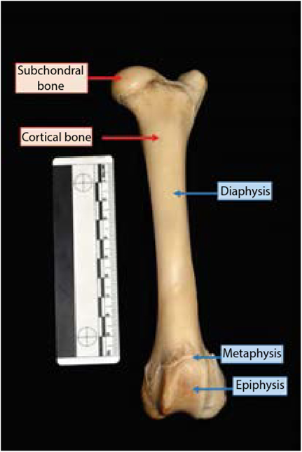
Figure 11.2 Parts of the bone and denoting compact versus subchondral bone.

Figure 11.3 Compact bone versus trabecular bone. Image of deer vertebra.

Figure 11.4 Mammal bone (deer) versus bird bone (turkey). Note the differences in the cortical bone thickness and the arrangement of the trabecular bone.
and then the species that are present. It also helps us understand the life histories of the individuals. Bone is built and modified according to Wolff’s law, which was outlined by an orthopedic surgeon, Julius Wolff (1892), and simply states that bone is laid down where it is needed and resorbed where it is not.
Wolff’s law is the foundation of the concept that form follows function; the skeleton of each class of animal (mammal, bird, reptile, and fish) has a generalized body plan with modifications that are grouped in class, genus, and species levels. These modifications are primarily based on locomotion (function) differences, that is, quadrupedal walking/running, slithering, flying, climbing, swinging, and so on, and dietary differences (i.e., herbivore, omnivore, carnivore).
Bones come in five basic forms, which help with initial identification. These are the long bones (limb bones; predominately compact bone), short bones (wrist and ankle bones; predominately spongy bone), flat bones (skull bones; plates of compact bone enclosing spongy bone), irregular bones (vertebrae; variable in composition), and sesamoid bones (patellae; bony structures that develop within tendons or ligaments).
Long bones are composed of three parts: the diaphysis (shaft), the epiphysis (end or joint surfaces), and the metaphysis (at the proximal and distal ends of the diaphysis) where bone growth occurs. During growth, a physis (growth plate) is present until the full length of the shaft is reached, at which point the diaphysis has met the epiphysis and fusion of the two begins. This process is tightly controlled by genetics and occurs in a highly predictable fashion, so as you can imagine, understanding the process could be useful for age determination (Figure 11.3).
Bones are not just straight tubes; they have bumps, ridges, and valleys, all of which have a name (see France, 2009 and White et al., 2012 for lists of features). These features are instrumental in determining the bones that are present, the side of the body from which the bone comes (i.e., right or left), and ultimately their class, genus, or species level association.

