Radiology and Imaging of the Osseous Response to Trauma
In forensic evaluations, radiographs are often utilized to estimate the chronicity of a fracture. The presence or absence of an osseous reaction to trauma predicts whether a skeletal injury occurred before death or postmortem. Identifying mild periosteal reaction associated with a fracture confirms that the fracture did not occur immediately prior to death and is not due to postmortem trauma. In the live animal, the radiographic changes identified in bone following trauma are variable based on the animal’s age, nutritional status or health, location of the injury, and degree of immobilization or weight-bearing status. In addition, most knowledge regarding the radiographic appearance of healing bone is based on clinical experience and on the study of fractures that have been stabilized or have undergone direct healing. With knowledge and understanding of these confounding factors, the radiographic progression of change in injured bone over time can be used as a general guideline to the timing of injuries (Figure 18.7a,b).
Bone is dynamic and changes during growth, following injury, and throughout life by a process of resorption and new bone production called remodeling. The radiographic identification of skeletal injury and the remodeling which follows injury are used in forensic evaluations. Radiographs can
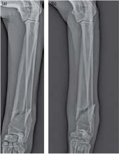
Figure 18.7 Craniocaudal projections of the antebrachium in a dog. (a) The sharp margins of the mildly comminuted distal antebrachial fracture are consistent with an acute fracture, (b) At 3 weeks following injury periosteal callus and resorption at the fracture margins are visible.
indicate the type of skeletal injury and potentially the mechanism of injury. Radiography and imaging can be useful in assessing the healing process and estimating when the injury occurred. Radiography and imaging studies may help distinguish antemortem and postmortem trauma and in some cases differentiate perimortem and postmortem injury.
Bone can be anatomically divided into regions of the periosteum or periosteal membrane, cortex, medullary cavity, nutrient foramen, endosteum, and articular surfaces. The periosteum can provide important evidence in medicolegal cases, such as how the trauma occurred, when it occurred, and if the injury is healing. The periosteum, which covers the entire bone with the exception of articular cartilage, can provide important components needed in bone healing and in bone growth of the immature animal. In the immature animal the periosteum is tightly attached to the underlying bone at the level of the physis and very loosely attached throughout the remainder of the bone. This configuration explains the appearance of the Salter-Harris II fracture, which cannot propagate completely across the physis due to the tight periosteal attachment, and deviates through the metaphysis (Figure 18.8). In the area of loose periosteal attachment, subperiosteal hemorrhage can occur secondary to trauma. The inner cambium layer of the periosteum is thick and cell-rich in the immature animal and gradually decreases in cellularity and thickness with age. This change in the structure of the periosteum explains why periosteal mineralization or periosteal reaction will occur earlier and will be more pronounced with trauma to immature bone compared to mature bone. Periosteal reactions are described as continuous or interrupted and by various descriptive terms such as solid, lamellated, palisading, spiculated, sunburst, and Codman triangle. Stable healing fractures show a smooth periosteal pattern. An onion skin or lamellated periosteal reaction may occur with waxing and waning disease or secondary to repeated trauma. Palisading periostitis can be seen with chronic soft tissue inflammation or with hypertrophic osteopathy. Spiculated new bone occurs with rapidly changing disease such as neoplasia. Sunburst patterns and Codman triangle are often associated with rapidly growing aggressive processes where the underlying disrupted periosteum has not had time to mineralize.
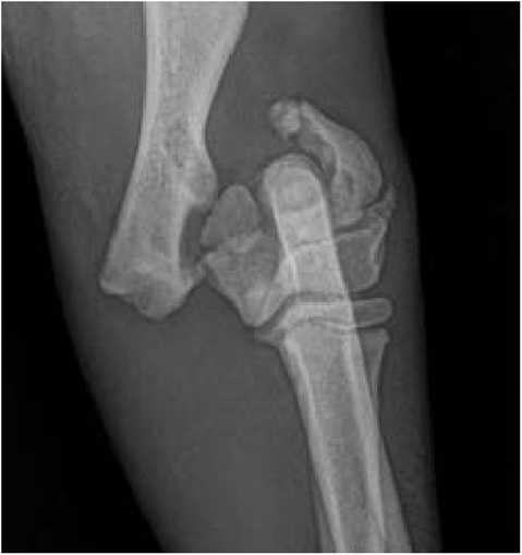
Figure 18.8 Craniocaudal view of the elbow in a young dog. The fracture has propagated through the medial distal humeral physis and exited through the lateral condyle. Note that the medial humeral epicondyle, which typically fuses to the remainder of the humerus between 6 and 8 months of age, is unfused. The medial and lateral condyles are fused.
Periostitis may be the first sign of accidental or nonaccidental trauma and may be present without a fracture and without outward signs of trauma. Torsional force may tear the periosteum in an immature animal and the secondary periosteal mineralization will be radiographically apparent days after injury. Periosteal mineralization can first be seen as early as 5 days or as late as 10 days following injury. Cases of suspected nonaccidental trauma with no abnormalities on the initial radiographic study can be radiographed in 10 days to look for early periosteal mineralization. Nondisplaced fractures that are occult on an initial radiographic examination may become conspicuous on follow-up imaging performed between 5 and 10 days, when the periosteal reaction is visible. The amount of periosteal reaction will progress with increased displacement or motion at a fracture site.
In addition to evaluation of the periosteum, assessment of the cortex, endosteum, medullary cavity, callus, and soft tissues at a fracture site may provide estimates as to the duration of fracture and trauma. In the 5 to 10 days following a fracture, the margins of fracture fragments will become less sharp and may decrease in opacity. In clinical imaging, when comparison images are available, an increased fracture gap may be initially identified due to this loss of opacity. In the 10 to 20 days following a fracture, endosteal callus may be visualized. Callus is initially hazy and less opaque than the cortex. Callus will become progressively more opaque 20 to 30 days after fracture. Identification of trabecular bone within a callus and remodeling generally take at least 30 days to occur.
Evaluation of the soft tissues at a fracture site may also provide some indication of the age of a fracture. Radiographically identified soft tissue swelling associated with a fracture will be pronounced in the first few days following injury and will diminish during the weeks after the trauma. The loss of soft tissues or soft tissue atrophy can be noted as early as 2 weeks following injury of the appendicular skeleton. When the contralateral limb is available for comparison, detecting marked muscle atrophy more likely indicates an injury of several weeks’ duration. Decreased bone opacity distal to a fracture site or osteopenia is further support of disuse and chronicity of a lesion.
Although radiography is primarily used for evaluating osseous trauma and is readily available in the veterinary field, other imaging modalities can assist in the forensic evaluation of osseous trauma. US by an experienced operator can detect cortical defects, callus, and hemorrhage. Removal of the air interface between the US probe and skin surface would require shaving the area of interest, and due to pain associated with the direct pressure of the transducer, sedation of the animal would likely be necessary. When available, CT and MR imaging are excellent adjunctive studies in the forensic evaluation of the bone’s response to trauma due to their superior contrast resolution and cross-sectional imaging capabilities and the ability of MR to detect areas of increased vascularity or bone marrow lesions (previously referred to as bone marrow edema). The potential of PMMR to provide evidence of a vital reaction could be useful in cause of death determinations.

